
Sebastian Stach
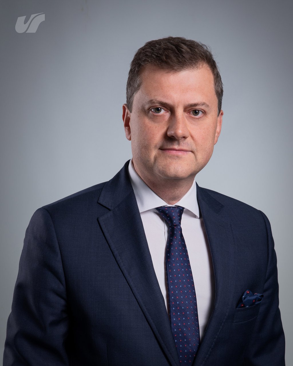
Dr hab. Sebastian Stach, prof. UŚ, jest dyrektorem Instytutu Inżynierii Biomedycznej Wydziału Nauk Ścisłych i Technicznych Uniwersytetu Śląskiego w Katowicach i przewodniczącym Rady Naukowej Instytutu. Funkcję dyrektora pełni od 2019 r., a w 2026 r. został powołany na kolejną kadencję, do 2030 r. Kieruje Laboratorium Neuroergonomii i Terapii Cyfrowych DriveLab, w którym rozwijane są symulatory jazdy i cyfrowe narzędzia wspierające rehabilitację oraz badania nad interakcją człowieka z technologią.
Z Uniwersytetem Śląskim związany jest od 1994 r., kiedy rozpoczął studia na Wydziale Techniki. Stopień doktora nauk technicznych w dyscyplinie inżynieria materiałowa uzyskał w 2005 r. na Wydziale Matematyki, Fizyki i Chemii UŚ. Stopień doktora habilitowanego nauk technicznych w dyscyplinie biocybernetyka i inżynieria biomedyczna uzyskał w 2017 r. w Instytucie Biocybernetyki i Inżynierii Biomedycznej im. Macieja Nałęcza PAN. W latach 2015-2019 był zastępcą dyrektora Instytutu Informatyki UŚ. Przez wiele lat kierował Laboratorium Ilościowej Analizy i Modelowania Powierzchni Biomateriałów ConLab. Był członkiem Senatu Uniwersytetu Śląskiego oraz Komitetu Biocybernetyki i Inżynierii Biomedycznej PAN.
Prowadzi badania z zakresu inżynierii biomedycznej i metrologii powierzchni. Zajmuje się ilościową analizą i modelowaniem powierzchni biomateriałów, analizą stereometryczną, fraktalną i multifraktalną, mikroskopią konfokalną i AFM, przetwarzaniem i analizą obrazów, informatyką biomedyczną oraz wytrzymałością materiałów. Jest opiekunem Studenckiego Koła Naukowego NeuroDrive.
Miejsce zatrudnienia
Uniwersytet Śląski w Katowicach, ul. Bankowa 12, 40-007 Katowice
Miejsce wykonywania pracy: Instytut Inżynierii Biomedycznej, Wydział Nauk Ścisłych i Technicznych, ul. Będzińska 39, 41-205 Sosnowiec
Aktualnie pełnione funkcje na uczelni
Dyrektor
Instytutu Inżynierii Biomedycznej
Przewodniczący
Rady Naukowej Instytutu Inżynierii Biomedycznej
Kierownik
Laboratorium Neuroergonomii i Terapii Cyfrowych DriveLab
Opiekun
Studenckiego Koła Naukowego NeuroDrive
Funkcje pełnione w przeszłości
Zastępca dyrektora
Instytutu Informatyki, 2015-2019
Kierownik
Laboratorium Ilościowej Analizy i Modelowania Powierzchni Biomateriałów ConLab, które utworzył z kierowanego przez siebie projektu FNiTP, 2011-2025
Członek
Senatu Uniwersytetu Śląskiego w Katowicach, kadencje 2016-2020 (w grupie młodszych pracowników nauki) i 2020-2024
Członek
Komitetu Biocybernetyki i Inżynierii Biomedycznej Polskiej Akademii Nauk, 2020-2023
Autor programu i kierownik
studiów podyplomowych „E-zdrowie”, od roku akademickiego 2014/2015 do zamknięcia studiów po drugim naborze
Przewodniczący
Kierunkowego Zespołu Zapewniania Jakości Kształcenia dla kierunku Inżynieria biomedyczna, 2013-2020
Kierownik zespołu
ds. opracowania programu kształcenia dla studiów II stopnia na kierunku Inżynieria biomedyczna (studia uruchomione w roku akademickim 2015/2016), 2015
Zastępca kierownika zespołu
ds. opracowania programu kształcenia kierunku Inżynieria biomedyczna według Krajowych Ram Kwalifikacji, autor większości dokumentacji, 2011-2012
Koordynator
Zintegrowanych Programów Uniwersytetu Śląskiego (POWER 3.5) w Instytucie Informatyki, 2018
Członek
Zespołu ds. wdrażania Strategii Uniwersytetu Śląskiego w Katowicach, 2019
Członek
Zespołu ds. Wdrażania Krajowych Ram Kwalifikacji, organu pomocniczego Rektora UŚ, od 2013
Członek
Komisji ds. studiów podyplomowych, organu pomocniczego Rektora UŚ, od 2014
Członek
Rady Programowej Centrum Kształcenia na Odległość UŚ jako przedstawiciel Instytutu Informatyki, od 2012
Członek
Rady Programowej kierunku Inżynieria biomedyczna (ciało kolegialne z udziałem przedstawicieli biznesu), od 2015
Opiekun
Studenckiego Koła Naukowego Inżynierii Biomedycznej InBio, 2015-2020
Opiekun roku
studiów I stopnia na kierunku Inżynieria biomedyczna (2010-2014, cały tok studiów) oraz studiów II stopnia (2016-2017)
Przebieg pracy zawodowej
(2019 - nadal)
profesor uczelni, Instytut Inżynierii Biomedycznej, Wydział Nauk Ścisłych i Technicznych
(2018 - 2019)
profesor uczelni, Zakład Komputerowych Systemów Biomedycznych, Instytut Informatyki, Wydział Informatyki i Nauki o Materiałach
(2009 - 2018)
adiunkt, Zakład Komputerowych Systemów Biomedycznych, Instytut Informatyki, Wydział Informatyki i Nauki o Materiałach
(2006 - 2009)
adiunkt, Zakład Badań Warstwy Wierzchniej, Katedra Materiałoznawstwa, Wydział Informatyki i Nauki o Materiałach
(2000 - 2006)
asystent, Zakład Badań Warstwy Wierzchniej, Katedra Materiałoznawstwa, Wydział Informatyki i Nauki o Materiałach
Wskaźniki bibliometryczne
PUBLIKACJE
ARTYKUŁY na liście JCR
sumaryczny IF
Średni IF na publikację z JCR
Sumaryczna punktacja ministerialna
kierowanych projektów i zadań
udziałów w projektach
patent
współpracujących jednostek naukowych
Zrealizowanych prac dyplomowych
zrealizowanych doktoratów
doktoratów w trakcie
indeks h Web of Science
indeks h Scopus
indeks h Google Scholar
Indeks h i liczba cytowań według baz, stan na 5 października 2026 r.:
- Web of Science Core Collection: 27 (1685 cytowań)
- Scopus: 27 (1799 cytowań)
- Google Scholar: 30 (2407 cytowań)
Wykształcenie
(2016 - 2017)
doktor habilitowany nauk technicznych w dyscyplinie biocybernetyka i inżynieria biomedyczna, Instytut Biocybernetyki i Inżynierii Biomedycznej im. Macieja Nałęcza Polskiej Akademii Nauk; osiągnięcie naukowe: „Modelowanie, symulacja komputerowa, ilościowa analiza i wizualizacja powierzchni biomateriałów na podstawie danych topograficznych uzyskanych za pomocą mikroskopii konfokalnej i mikroskopii AFM”; stopień nadany 25.04.2017
(1999 - 2005)
doktor nauk technicznych w dyscyplinie inżynieria materiałowa, studia doktoranckie na Wydziale Matematyki, Fizyki i Chemii Uniwersytetu Śląskiego w Katowicach; rozprawa wyróżniona: „Zastosowanie stereometrii powierzchni i analizy multifraktalnej w opisie morfologii przełomu materiałów” (Wydział Informatyki i Nauki o Materiałach UŚ, 2005)
(1994 - 1999)
magister techniki, jednolite studia magisterskie na Wydziale Techniki Uniwersytetu Śląskiego w Katowicach, kierunek: Wychowanie Techniczne, specjalność: Technika i Informatyka; praca magisterska: „Fraktalny opis struktury pasmowej”
Projekty badawcze
Kierowanie projektami
- Nieinwazyjny system nadzoru parametrów fizjologicznych sportowców w oparciu o rejestrację wielkości stanowiących złoty standard oceny efektywności treningu, w warunkach rzeczywistych, 2024-2028, Doktorat wdrożeniowy (MNiSW), kierownik projektu
- Nieuciążliwy system monitorowania i analizy epizodów kaszlu w diagnostyce chorób układu oddechowego w szczególności astmy z zastosowaniem algorytmów sztucznej inteligencji w systemach wbudowanych, 2024-2028, Doktorat wdrożeniowy (MNiSW), kierownik projektu
- Opracowanie przestrzennego modelu materiału, 2018-2019, Narodowe Centrum Nauki, MINIATURA, kierownik projektu
- Wyposażenie laboratorium ilościowej analizy i modelowania powierzchni biomateriałów w pomiarowy skaningowy laserowy mikroskop konfokalny, 2010-2012, 599/FNITP/160/2010, autor projektu, kierownik i główny wykonawca
- Rozwój metod ilościowego opisu mikrostruktury i powierzchni materiałów, Podtemat: Komplementarne zastosowanie parametrów multifraktalnych, transformaty falkowej i perkolacji w opisie powierzchni przełomu materiału, 2009, BW-08-0500-017-09, kierownik projektu i wykonawca podtematu
- Transformata falkowa - analiza zagadnienia, 2008, BW/KM/338/2008, badania własne, kierownik projektu
- Współczesne systemy badania topografii powierzchni. Określenie możliwości i porównywalności zapisu obrazów 3D. Etap 1, 2007, BW/KM/209 zad. 3/2007, badania własne, kierownik projektu
Kierowanie zadaniami w projektach
- Zintegrowany Program 3.5 POWER, 2018-2023, POWR.03.05.00-IP.08-00-PZ1/17, Program Operacyjny Wiedza Edukacja Rozwój, kierownik zadania
- Jeden Uniwersytet - Wiele Możliwości. Program Zintegrowany, 2019-2023, Program Operacyjny Wiedza Edukacja Rozwój, kierownik zadania
Udział w projektach jako wykonawca
- Computational modelling of biomechanics in the musculoskeletal system: tissues, replacements and regeneration, Podtemat: Modelling fracture processes in orthopaedic implants, 2011-2014, Projekt międzynarodowy, wykonawca podtematu
- Opracowanie nowych gatunków supergruboziarnistych węglików spiekanych z osnową zawierającą nikiel, 2012-2013, N507 222240, Politechnika Śląska, wykonawca badań
- Badania statutowe Zakładu Badań Warstwy Wierzchniej Katedry Materiałoznawstwa UŚ, 2001-2008, BS/KM/209, wykonawca zadań
- Opracowanie algorytmu charakteryzującego stereometrycznie powierzchnię na podstawie analizy głębi ostrości z mikroskopu świetlnego, 2006, BW/KM/209 zad. 3/2006, badania własne, wykonawca zadania
- Wpływ czynników strukturalnych na właściwości ceramiki sialonowej w ujęciu metod materiałografii i fraktografii ilościowej, 2004-2006, 4T08D 01822, wykonawca badań
- Ilościowy opis morfologii powierzchni przełomu tradycyjną metodą fraktografii, 2004, BW/KM/209 zad. 1/2004, badania własne, wykonawca zadania
- Stereometryczny i fraktalny opis powierzchni przełomu materiałów, 2003, BW/KM/209 zad. 3/2003, badania własne, wykonawca zadania
- Weryfikacja koncepcji opisu morfologii przełomu metodami fraktografii ilościowej i analizy multifraktalnej, 2002, BW/KM/209 zad. 6/2002, badania własne, wykonawca zadania
Udział naukowy w projektach finansujących badania (według podziękowań w publikacjach)
- Fundusze Europejskie dla Śląska 2021-2027 ()
- Narodowe Centrum Nauki, SONATA, nr 2011/03/D/ST8/04884 ()
- Narodowe Centrum Nauki, PRELUDIUM, nr 2016/23/N/ST8/03809 ()
- Narodowe Centrum Nauki, PRELUDIUM, nr 2011/01/N/NZ5/00919 ()
- MNiSW, Diamentowy Grant, nr DI2015 019045 ()
- MNiSW, Inkubator Innowacyjności+, nr 10/NAB3/II+/2017 ()
- MNiSW, grant nr N N402 471337 ()
- Komitet Badań Naukowych, granty na eksperyment ICARUS ()
- UEFISCDI (Rumunia), nr PN-II-PT-PCCA-2013-4-1232 ()
- Ministerstwo Edukacji, Nauki i Rozwoju Technologicznego Republiki Serbii, projekt nr 172003 ()
- Ministerstwo Edukacji, Nauki i Rozwoju Technologicznego Republiki Serbii, projekt nr TR 035020 ()
- Ministerstwo Edukacji, Nauki i Rozwoju Technologicznego Republiki Serbii, projekt nr III-45006 ()
- European Centre of Excellence CEITEC, nr CZ.1.05/1.1.00/02.0068 ()
- Sensor, Information and Communication Systems SIX, nr CZ.1.05/2.1.00/03.0072 ()
- Grant FEKT-S-14-2240 (Politechnika w Brnie) ()
- Grant FEKT-S-17-4626 (Politechnika w Brnie) ()
- CEITEC 2020, nr LQ1601 ()
- National Sustainability Programme, nr LO1401 ()
- CEITEC Nano Research Infrastructure, nr LM2015041 ()
- Czech Science Foundation (GAČR), nr 15-05259S ()
- Visegrad Fund ()
- State assignments nr 2560 i nr 16.1103.2014K ()
- Ministry of Education, Youth and Sports of the Czech Republic, projekt nr CZ.1.07/2.3.00/30.0021 Strengthening of Research and Development Teams at the University of Pardubice ()
- Bulgarian National Science Fund, projekt nr DO 02-129/08 ()
- Universiti Sains Malaysia, grant nr 1001/PFIZIK/843088 ()
Współpraca z ośrodkami naukowymi w kraju i za granicą
- Akademia Górniczo-Hutnicza im. Stanisława Staszica w Krakowie, Wydział Energetyki i Paliw, al. Mickiewicza, Kraków ()
- Akademia Górniczo-Hutnicza im. Stanisława Staszica w Krakowie, Wydział Fizyki i Informatyki Stosowanej, Katedra Fizyki Medycznej i Biofizyki, al. Mickiewicza, Kraków ()
- Akademia Górniczo-Hutnicza im. Stanisława Staszica w Krakowie, Wydział Odlewnictwa, ul. Reymonta 23, 30-059 Kraków ()
- Akademia Wychowania Fizycznego im. Jerzego Kukuczki w Katowicach, Instytut Nauk o Sporcie, ul. Mikołowska 72A, 40-065 Katowice ()
- Akademia Wychowania Fizycznego im. Jerzego Kukuczki w Katowicach, Wydział Wychowania Fizycznego, Katedra Treningu i Żywienia w Sporcie, ul. Mikołowska 72A, 40-065 Katowice ()
- Akademia Wychowania Fizycznego im. Jerzego Kukuczki w Katowicach, Wydział Zarządzania Sportem i Turystyką, ul. Mikołowska 72A, 40-065 Katowice ()
- Alzahra University, Department of Physics, Tehran, Iran ()
- Arak University of Technology, Department of Mechanical Engineering, Arak, Iran ()
- Brno University of Technology, Central European Institute of Technology (CEITEC), Purkyňova 123, 612 00 Brno, Czech Republic ()
- Brno University of Technology, Faculty of Electrical Engineering and Communication, Physics Department, Technická 8, 616 00 , Brno, Czech Republic ()
- Center of Research and Technological Development in Electrochemistry (CIDETEQ), Parque Tecnológico Sanfandila, Pedro Escobedo, 76703, Santiago de Querétaro, Mexico ()
- Ciudad Universitaria Puebla, Benemérita Universidad Autónoma de Puebla, Chemistry Center ICUAP, 72530, Puebla, Mexico ()
- Dagestan State University, Faculty of Physics, st. M. Gadjieva 43-a, 367015, Makhachkala, Dagestan Republic, Russia ()
- Dental Polyclinic, Perkovceva 3, 10 000 , Zagreb, Croatia ()
- Guru Nanak Dev University, Department of Physics, Material Science Research Laboratory, Amritsar, 143005, Punjab, India ()
- Institut "Rudjer Boskovic", Bijenicka Cesta 54, 10000, Zagreb, Croatia ()
- Institut National de la Recherche Scientifique (INRS), 1650 Boulevard Lionel-Boulet, QC J3X 1P7, Varennes, Canada ()
- Islamic Azad University, Arak Branch, Young Researchers and Elite Club, Arak, Iran ()
- Islamic Azad University, Kermanshah Branch, Department of Physics, Kermanshah, Iran ()
- Islamic Azad University, Kermanshah Branch, Young Researchers and Elite Club, Kermanshah, Iran ()
- Islamic Azad University, Science and Research Branch, Department of Physics, Tehran, Iran ()
- Islamic Azad University, Science and Research Branch, Plasma Physics Research Center , P.O. Box 14665-678, Tehran, Iran ()
- Islamic Azad University, West Tehran Branch, Department of Physics, Tehran, Iran ()
- Islamic Azad University, West Tehran Branch, Young Researchers and Elite Club, Tehran, Iran ()
- Istituto Italiano di Tecnologia, Materials Characterization Facility, Via Morego 30, I-16163 Genova, Italy ()
- Istituto Italiano di Tecnologia, Nanophysics Department, Scanning Probe Microscopy Lab, Via Morego 30, I-16163, Genova, Italy ()
- "Iuliu Haţieganu" University of Medicine and Pharmacy, Faculty of Dentistry, Department of Periodontology, 8 Victor Babes St., 400012, Cluj-Napoca, Romania ()
- "Iuliu Haţieganu" University of Medicine and Pharmacy, Faculty of Medicine, Department of Surgical Specialties and Medical Imaging, Discipline of Ophthalmology, 8 Victor Babeş St., 400012, Cluj-Napoca, Romania ()
- Malayer University, Faculty of Science, Department of Physics, Malayer, Iran ()
- Medical University, Faculty of Pharmacy, Department of Organic Chemistry, Sofia, Bulgaria ()
- Nano Mabna Iranian Inc., P O Box 1676664116, Tehran, Iran ()
- Nuclear Sciences and Technology Research Institute, Physics and Accelerators Research School, Tehran, Iran ()
- Payame Noor University (PNU), Department of Physics, Tehran, Iran ()
- Payame Noor University, Department of Physics, P.O. Box 19395-4697, Tehran, Iran ()
- Politechnika Rzeszowska im. Ignacego Łukasiewicza, Wydział Budowy Maszyn i Lotnictwa, Katedra Nauki o Materiałach, 35-959 Rzeszów ()
- Politechnika Śląska, Instytut Fizyki - Centrum Naukowo-Dydaktyczne, ul. Konarskiego 22B, 44-100 Gliwice ()
- Politechnika Śląska, Wydział Automatyki, Elektroniki i Informatyki, ul. Akademicka 16, 44-100 Gliwice ()
- Politechnika Śląska, Wydział Inżynierii Biomedycznej, Katedra Biomateriałów i Inżynierii Wyrobów Medycznych, ul. Roosevelta 40, 41-800 Zabrze ()
- Politechnika Śląska, Wydział Inżynierii Materiałowej i Cyfryzacji Przemysłu (dawniej: Wydział Inżynierii Materiałowej i Metalurgii, Katedra Nauki o Materiałach; Wydział Inżynierii Materiałowej), ul. Krasińskiego 8, 40-019 Katowice ()
- Politechnika Śląska, Wydział Inżynierii Materiałowej i Cyfryzacji Przemysłu, Katedra Technologii Materiałowych, ul. Krasińskiego 8, 40-019 Katowice ()
- Politechnika Wrocławska, Wydział Budownictwa Lądowego i Wodnego, Wrocław ()
- Politechnika Wrocławska, Wydział Mechaniczny, Wrocław ()
- Promobil s.c., ul. Kopernika 12, 40-064 Katowice ()
- Queen's University, Department of Physics, Engineering Physics and Astronomy, Kingston, Ontario K7L 3N6, Canada ()
- Razi University, Faculty of Science, Department of Physics, Kermanshah, Iran ()
- Razi University, Nano Science and Technology Research Center, Kermanshah, Iran ()
- SICLAB Limited Liability Company, st. M. Yaragskogo 75, 367030, Makhachkala, Dagestan Republic, Russia ()
- Sieć Badawcza Łukasiewicz - Instytut Ceramiki i Materiałów Budowlanych, ul. Cementowa 8, 31-983 Kraków ()
- Sieć Badawcza Łukasiewicz - Instytut Materiałów Polimerowych (dawniej: Instytut Inżynierii Materiałów Polimerowych i Barwników), ul. Marii Skłodowskiej-Curie 55, 87-100 Toruń ()
- Sieć Badawcza Łukasiewicz - Instytut Metali Nieżelaznych, ul. Sowińskiego 5, 44-100 Gliwice ()
- Śląski Uniwersytet Medyczny w Katowicach, Wydział Nauk Medycznych w Katowicach, I Katedra Kardiologii, 40-055 Katowice ()
- Śląski Uniwersytet Medyczny w Katowicach, Wydział Nauk Medycznych w Katowicach, Katedra i Klinika Chorób Wewnętrznych, Autoimmunologicznych i Diabetologii (dawniej: Katedra i Klinika Chorób Wewnętrznych, Autoimmunologicznych i Metabolicznych), ul. Medyków 14, 40-572 Katowice ()
- Śląski Uniwersytet Medyczny w Katowicach, Wydział Nauk Medycznych w Katowicach, Katedra i Klinika Laryngologii, ul. Francuska 20/24, 40-027 Katowice ()
- Śląski Uniwersytet Medyczny w Katowicach, Wydział Nauk Medycznych w Katowicach, Katedra i Klinika Pneumonologii, 40-055 Katowice ()
- Śląski Uniwersytet Medyczny w Katowicach, Wydział Nauk Medycznych w Katowicach, Katedra i Zakład Epidemiologii, ul. Medyków 18, 40-752 Katowice ()
- Technical University of Cluj-Napoca, Faculty of Automotive, Mechatronics and Mechanical Engineering (dawniej: Faculty of Mechanical Engineering), Department of Automotive Engineering and Transportation, Discipline of Descriptive Geometry and Engineering Graphics, 103-105 B-dul Muncii St., 400641, Cluj-Napoca, Romania ()
- Technical University of Cluj-Napoca, The Directorate of Research, Development and Innovation Management (DMCDI), Constantin Daicoviviu Street, no. 15, 400020, Cluj-Napoca, Romania ()
- Universidad Autonoma de Nuevo Leon, Facultad de Ciencias Físico Matematicas, Av. Pedro de Alba s/n, San Nicolas de los Garza, , 66455, Nuevo Leon, Mexico ()
- Universidad Autónoma de Nuevo León (UANL), Facultad de Ciencias Físico Matemáticas (FCFM), Av. Universidad s/n, San Nicolás de los Garza, Nuevo León, Mexico ()
- Universiti Sains Malaysia, School of Physics, Nano-Optoelectronics Research and Technology Laboratory, 11800, Penang, Malaysia ()
- University of Belgrade, Vinča Institute of Nuclear Sciences, Mike Alasa 12-14, 11001, Belgrade, Serbia ()
- University of Bu Ali Sina, Department of Physics, P.O. Box 65174, Hamedan, Iran ()
- University of Craiova, Faculty of Mechanical Engineering, Department of Applied Mechanics, Calea Bucureşti St., no. 165, 200585, Craiova, Dolj, Romania ()
- University of Hradec Kralove, Department of Physics, Rokitansk eho 62, 500-03, Hradec Kralove, Czech Republic ()
- University of Miami, College of Engineering, Biomedical Atomic Force Microscopy Laboratory, McArthur Annex Room 170A, 1251 Memorial Drive, FL 33146, Coral Gables, USA ()
- University of Milano Bicocca, Department of Materials Science and COMiB Research Center, Milano, Italy ()
- University of Milano Bicocca, Department of Materials Science, Piazza dell’Ateneo Nuovo, 20125, Milano, Italy ()
- University of Namur, Research Centre in Physics of Matter and Radiation (PMR), LISE Laboratory, B-5000, Namur, Belgium ()
- University of Novi Sad, Faculty of Medicine, Department of Restorative Dentistry and Endodontics, Clinic of Dentistry of Vojvodina, Hajduk Veljkova 3, 21000, Novi Sad, Serbia ()
- University of Novi Sad, Faculty of Medicine, Department of Restorative Dentistry and Endodontics, Clinic of Dentistry of Vojvodina, School of Dentistry, Novi Sad, Serbia ()
- University of Novi Sad, Faculty of Medicine, School of Dentistry, Hajduk Veljkova 3, 21000, Novi Sad, Serbia ()
- University of Novi Sad, Faculty of Technical Sciences, Department for Production Engineering, Trg Dositeja Obradovica 6, 21000, Novi Sad, Serbia ()
- University of Palermo, Department of Mathematics and Informatics, Via Archirafi 34, 90123, Palermo, Italy ()
- University of Pardubice, Faculty of Chemical Technology and Centre for Material Science, Department of General and Inorganic Chemistry, Studentská 573, 532 10, Pardubice II, Czech Republic ()
- University of Raparin, Department of Physics, College of Science, Iraq ()
- University of Sofia, Faculty of Chemistry and Pharmacy, Department of Inorganic Chemistry, Sofia, Bulgaria ()
- University of Thi-Qar, Physics Department, Science College, Nassiriya Nanotechnology Research Laboratory (NNRL), 00964, Nassiriya, Iraq ()
- University of Zagreb, Faculty of Metallurgy, Aleja narodnih heroja 3, 44000 , Sisak, Croatia ()
- University of Zagreb, School of Dental Medicine, Gunduliceva 5, 10000 , Zagreb, Croatia ()
- Uniwersytet Jagielloński Collegium Medicum, Katedra Chorób Wewnętrznych i Gerontologii, ul. Śniadeckich, Kraków ()
- Uniwersytet Jagielloński, Wydział Biochemii, Biofizyki i Biotechnologii, 30-387 Kraków ()
- Vacuum Technology Group, ACECR - Sharif Branch, Tehran, Iran ()
- World Aquatics, CH-1005 Lausanne, Switzerland ()
Publikacje
- Gabor J, Roczniok R, Mikrut G, Szewczenko J, Popczyk M, Wilk K, Stach S, Karpati G, Mizia-Stec K, Kłeczek AM, Swinarew AS. Influence of Contact Angle and Wetting Angle on Water Polo Ball Performance: A Continuation Study. Applied Sciences. 2026;16(13):6686. doi:10.3390/app16136686 100 pkt13 IF 2,900 IF5 3,000
- Cebulski J, Pasek D, Roskosz S, Popczyk M, Gabor J, Stach S, Wrzalik R, Wojtyniak M, Simlot M, Swinarew AS. Topography and Corrosion Resistance Characteristics of Fe40Al5Cr0.2ZrB Alloy and X18CrN28 Steel. Materials. 2025;18(23):5465. doi:10.3390/ma18235465 140 pkt13 IF 3,700 IF5 3,800
- Piątkowski J, Roskosz S, Stach S, Górny M. Crystallisation and Microstructure of Sludge Particles in AlSi7Mg Secondary Alloys with Increased Iron Content. Materials. 2025;18(21):4921. doi:10.3390/ma18214921 140 pkt13 IF 3,700 IF5 3,800
- Piątkowski J, Roskosz S, Sapota W, Stach S. The Influence of Iron Content on the Porosity of AlSi9 Alloy Intended for Alfining Piston Ring Inserts. Materials. 2024;17(21):5181. doi:10.3390/ma17215181 140 pkt13 IF 3,200 IF5 3,500
- Piątkowski J, Roskosz S, Stach S. The Influence of Selected High - Pressure Die Casting Parameters on the Porosity of EN AB-46000 Alloy Castings. Advances in Science and Technology Research Journal. 2024;18(5):361-371. doi:10.12913/22998624/191236 100 pkt13 IF 1,300 IF5 1,100
- Gabor J, Mikrut G, Flak T, Cebo P, Roczniok R, Swinarew B, Langer E, Popczyk M, Stanula A, Stach S, Swinarew AS. Influence of Surface Structure on Ball Properties during a Professional Water Polo Game. Materials. 2023;16(8):3048. doi:10.3390/ma16083048 140 pkt12 IF 3,100 IF5 3,400
- Niedbała J, Popczyk M, Hawełek Ł, Orda S, Okła H, Gabor J, Stach S, Swinarew AS. Production of Electrolytic Composite Powder by Nickel Plating of Shredded Polyurethane Foam. Materials. 2022;15(11):3895. doi:10.3390/ma15113895 140 pkt11 IF 3,400 IF5 3,800
- Swinarew AS, Flak T, Jarosińska A, Garczyk Ż, Gabor J, Skoczyński S, Brożek G, Paluch J, Popczyk M, Stanula A, Stach S. Polyurethane-Based Porous Carbons Suitable for Medical Application. Materials. 2022;15(9):3313. doi:10.3390/ma15093313 140 pkt11 IF 3,400 IF5 3,800
- Garczyk Ż, Stach S. Three-Dimensional Model for Assessing the Pore Volume of Biomaterials Intended for Implantation. W: Computational Modelling of Biomechanics and Biotribology in the Musculoskeletal System: Biomaterials and Tissues (Second Edition). Woodhead Publishing Series in Biomaterials. Woodhead Publishing; 2021. s. 305-358. doi:10.1016/B978-0-12-819531-4.00013-4 20 pkt14 IF5 4,042
- Dercz G, Barczyk J, Matuła I, Kubaszek T, Góral M, Maszybrocka J, Bochenek D, Stach S, Szklarska M, Ryszawy D, Pudełek M. Characterization of YSZ Coatings Deposited on cp-Ti Using the PS-PVD Method for Medical Applications. Coatings. 2021;11(11):1348. doi:10.3390/coatings11111348 100 pkt11 IF 3,236 IF5 3,312
- Garczyk Ż, Jaegermann Z, Duda P, Swinarew AS, Stach S. Ceramic Biomaterial Pores Stereology Analysis by the Use of Microtomography. Materials. 2021;14(9):2207. doi:10.3390/ma14092207 140 pkt11 IF 3,748 IF5 4,042
- Łosiewicz B, Osak P, Maszybrocka J, Kubisztal J, Stach S. Effect of Autoclaving Time on Corrosion Resistance of Sandblasted Ti G4 in Artificial Saliva. Materials. 2020;13(18):4154. doi:10.3390/ma13184154 140 pkt11 IF 3,623 IF5 3,920
- Sapota W, Szczepanik P, Stach S, Wróbel Z. Fractal and Multifractal Analyses of the Porosity Degree of Ceramics Used in Biomedicine. Advanced Science, Engineering and Medicine. 2020;12(4):450-456. doi:10.1166/asem.2020.2546 20 pkt11
- Stach S, Ţălu Ş, Dallaev R, Arman A, Sobola D, Salerno M. Evaluation of the Topographical Surface Changes of Silicon Wafers after Annealing and Plasma Cleaning. Silicon. 2020;12(11):2563-2570. doi:10.1007/s12633-019-00351-x 40 pkt11 IF 2,670 IF5 2,474
- Szklarska M, Łosiewicz B, Dercz G, Maszybrocka J, Rams-Baron M, Stach S. Electrophoretic deposition of chitosan coatings on the Ti15Mo biomedical alloy from a citric acid solution. RSC Advances. 2020;10(23):13386-13393. doi:10.1039/d0ra01481h 100 pkt11 IF 3,361 IF5 3,390
- Ţălu Ş, Rąplewicz A, Stach S, Ţălu M, Vintilă D. 3-D Morphological Analysis of Carbon-Nickel Nanocomposite Thin Films. Applied Mechanics and Materials. 2020;896:311-320. doi:10.4028/www.scientific.net/AMM.896.311 5 pkt19
- Brożyna M, Stach S, Wróbel Z. Rozwój telemedycyny w Polsce po wdrożeniu „Elektronicznej Platformy Gromadzenia, Analizy i Udostępniania zasobów cyfrowych o Zdarzeniach Medycznych (P1)”. W: Lipowicz I, Szpor G, Świerczyński M, red. Telemedycyna i e-zdrowie. Prawo i informatyka. 2019. s. 89-104. 20 pkt14
- Garczyk ŻA, Stach S, Wróbel Z. Evaluation of Implant Surface Porosity Using IT Tools. W: The Book of Articles National Scientific Conference “Science and Young Researchers, III Edition - June 15, 2019, Łódź. Promovendi Foundation Publishing; 2019. s. 59-66. 5 pkt14
- Garczyk Ż, Stach S, Tălu Ş, Sobola D, Wróbel Z. Segmentation of Three-Dimensional Images of the Butterfly Wing Surface. W: Pietka E, Badura P, Kawa J, Wieclawek W, red. Information Technology in Biomedicine. ITIB 2018. Advances in Intelligent Systems and Computing, Vol. 762. Cham: Springer; 2019. s. 111-121. doi:10.1007/978-3-319-91211-0_10 20 pkt14
- Płatek M, Sapota W, Stach S, Wróbel Z. Reconstruction of Gigapixel Stereometric Maps of Ceramic Surfaces. W: Pietka E, Badura P, Kawa J, Wieclawek W, red. Information Technology in Biomedicine. ITIB 2018. Advances in Intelligent Systems and Computing, Vol. 762. Cham: Springer; 2019. s. 101-110. doi:10.1007/978-3-319-91211-0_9 20 pkt14
- Sapota WA, Stach S, Wróbel Z. Devices Supporting Gait Reeducation. W: The Book of Articles National Scientific Conference “Science and Young Researchers, III Edition - June 15, 2019, Łódź. Promovendi Foundation Publishing; 2019. s. 118-132. 5 pkt14
- Dejam L, Solaymani S, Achour A, Stach S, Ţălu Ş, Beryani Nezafat N, Dalouji V, Shokri AA, Ghaderi A. Correlation between surface topography, optical band gaps and crystalline properties of engineered AZO and CAZO thin films. Chemical Physics Letters. 2019;719:78-90. doi:10.1016/j.cplett.2019.01.042 70 pkt11 IF 2,029 IF5 1,724
- Dercz GS, Matuła IM, Maszybrocka J, Zubko MJ, Barczyk J, Pająk L, Stach S. Effect of Milling Time and Presence of Sn on the Microstructure and Porosity of Sintered Ti-10Ta-8Mo and Ti-10Ta-8Mo-3Sn Alloys. Journal of Alloys and Compounds. 2019;791:232-247. doi:10.1016/j.jallcom.2019.03.287 100 pkt11 IF 4,650 IF5 4,082
- Dallaev R, Stach S, Ţălu Ş, Sobola D, Méndez-Albores A, Trejo Córdova G, Grmela L. Stereometric Analysis of Effects of Heat Stressing on Micromorphology of Si Single Crystals. Silicon. 2019;11(6):2945-2959. doi:10.1007/s12633-019-0085-4 40 pkt11 IF 1,499 IF5 1,444
- Stach S, Ţălu Ş, Abdolghaderi S, Shafiekhani A, Bahmani J. 3_D surface stereometry of Ag/DLC nanocomposite prepared by RF-PECVD. Results in Physics. 2019;15:102731. doi:10.1016/j.rinp.2019.102731 70 pkt11 IF 4,019 IF5 3,706
- Stach S, Ţălu Ş, Głuchaczka A, Siek P, Zając J, Tavazzi S. Microscopic investigations of surface texture of siloxane-hydrogel contact lenses. Polymer Engineering and Science. 2019;59(S2):E442-E451. doi:10.1002/pen.25080 70 pkt11 IF 1,917 IF5 1,846
- Stach S, Kędzia O, Garczyk Ż, Wróbel Z. Modelling the degree of porosity of the ceramic surface intended for implants. Biomedizinische Technik. 2019;64(2):215-223. doi:10.1515/bmt-2017-0169 40 pkt11 IF 1,054 IF5 0,415
- Ţălu Ş, Stach S, Lainović T, Blažić L. Characterization of spatial patterns of dental restorative nanocomposites. Microscopy Research and Technique. 2019;82(7):1215-1223. doi:10.1002/jemt.23270 70 pkt11 IF 2,117 IF5 1,663
- Ţălu Ş, Stach S, Klaić B, Čelebić A. Evaluation of Topographical Co-Cr-Mo Alloy Surface Changes After Various Finishing Treatments. Acta Stomatologica Croatica. 2019;53(3):264-273. doi:10.15644/asc53/3/8 20 pkt11
- Ţălu Ş, Stach S, Abdolghaderi S. The effects of deposition time on the nanoscale patterns of Ag/DLC nanocomposite synthesized by RF-PECVD. Microscopy Research and Technique. 2019;82(5):572-579. doi:10.1002/jemt.23203 70 pkt11 IF 2,117 IF5 1,663
- Garczyk Ż, Stach S, Wróbel Z. Przetwarzanie i analiza obrazu w ocenie stopnia porowatości powierzchni bioceramiki. W: Czyż Z, red. Współczesne problemy inżynierii materiałowej oraz mechaniki. Lublin: Wydawnictwo Naukowe TYGIEL; 2018. s. 105-115. 20 pkt14
- Garczyk ŻA, Stach S, Wróbel Z. Przetwarzanie i analiza obrazu w ocenie stopnia porowatości powierzchni bioceramiki : [prezentacja ustna]. W: X Interdyscyplinarna Konferencja Naukowa Tygiel 2018 „Interdyscyplinarność kluczem do rozwoju” Lublin, 17-18 marca 2018 r. abstrakty. Fundacja na rzecz Promocji Nauki i Rozwoju Tygiel; 2018. s. 181-182.
- Sapota WA, Stach S, Wróbel Z. Możliwości oceny powierzchni biomateriałów z wykorzystaniem komercyjnych rozwiązań przetwarzania i analizy obrazu : [prezentacja ustna]. W: X Interdyscyplinarna Konferencja Naukowa Tygiel 2018 „Interdyscyplinarność kluczem do rozwoju” Lublin, 17-18 marca 2018 r. abstrakty. Fundacja na rzecz Promocji Nauki i Rozwoju Tygiel; 2018. s. 172.
- Sapota W, Stach S, Wróbel Z. Możliwości oceny powierzchni biomateriałów z wykorzystaniem komercyjnych rozwiązań przetwarzania i analizy. W: Czyż Z, red. Współczesne problemy inżynierii materiałowej oraz mechaniki. Lublin: Wydawnictwo Naukowe TYGIEL; 2018. s. 91-104. 20 pkt14
- Konsek D, Stach S, Ţălu Ş, Naderi S, Arman A. Correlation Between 3-D Surface Topography and Different Deposition Times of Engineered Ni@a-C:H Thin Films. Silicon. 2018;10(5):2141-2151. doi:10.1007/s12633-017-9743-6 25 pkt10 IF 1,210 IF5 1,281
- Kubisztal J, Kubisztal M, Stach S, Haneczok G. Corrosion resistance of anodic coatings studied by scanning microscopy and electrochemical methods. Surface and Coatings Technology. 2018;350:419-427. doi:10.1016/j.surfcoat.2018.07.032 35 pkt10 IF 3,192 IF5 3,110
- Garczyk Ż, Stach S, Wróbel Z. Analiza trójwymiarowych obrazów w badaniach topografii powierzchni materiału. W: Czyż Z, Maciąg K, red. Rozwój tworzyw inżynierskich i nauk o materiałach. Lublin: Wydawnictwo Naukowe TYGIEL; 2017. s. 16-29. 20 pkt14
- Garczyk Ż, Stach S, Wróbel Z. Analiza trójwymiarowych obrazów w badaniach topografii powierzchni materiału [abstrakt]. W: Nowak BA, Maciąg M, Maciąg K, red. IX Interdyscyplinarna Konferencja Naukowa TYGIEL 2017 „Interdyscyplinarność kluczem do rozwoju”. Abstrakty; 2017 Mar 18-19; Lublin. Lublin: Fundacja na rzecz promocji nauki i rozwoju TYGIEL; 2017. s. 431.
- Sapota WA, Stach S, Wróbel Z. Możliwości programu Matlab w zakresie analizy i wizualizacji powierzchni zespoleń kostnych stosowanych w leczeniu urazów twarzoczaszki. W: Aplikacyjne metody obliczeniowe oraz zarządzanie danymi. 2017. s. 198-214. 20 pkt14
- Sapota W, Stach S, Wróbel Z. Możliwości programu Matlab w zakresie analizy i wizualizacji powierzchni zespoleń kostnych stosowanych w leczeniu urazów twarzoczaszki [abstrakt]. W: Nowak BA, Maciąg M, Maciąg K, red. IX Interdyscyplinarna Konferencja Naukowa TYGIEL 2017 „Interdyscyplinarność kluczem do rozwoju”. Abstrakty; 2017 Mar 18-19; Lublin. Lublin: Fundacja na rzecz promocji nauki i rozwoju TYGIEL; 2017. s. 420-421.
- Ţălu Ş, Lainovic T, Stach S, Vilotić M, Blažić L. Analysis of the Multifractal Intrinsic Nature of Dental Nanocomposites’ Surface [Abstract]. W: 12th Conference for Young Scientists in Ceramics (CYSC-2017), October 18th-21st, Novi Sad, Serbia. Abstract Book. 2017. s. 53.
- Ţălu Ş, Sobola D, Tománek P, Stach S. Micromorphology of AlN Epilayers on Sapphire Substrates. W: International Conference on Computer, Electronics and Communication Engineering (CECE 2017); 2017 Jun 25-26; Sanya, China. DEStech Transactions on Computer Science and Engineering. DEStech Publications; 2017. s. 465-470. doi:10.12783/dtcse/cece2017/14582 5 pkt14
- Adamiec A, Stach S, Ţălu Ş. Quantitative Investigations About the Surface Texture Characteristics of the Nickel-Carbon Composite Thin Films Using Stereometric Analysis. W: Proceedings of the 2017 International Conference on Applied Mathematics, Modeling and Simulation (AMMS 2017); 2017 Nov 26-27; Shanghai, China. Vol. 153. Paris: Atlantis Press; 2017. s. 98-101. doi:10.2991/amms-17.2017.22 15 pkt16
- Gajos-Grzetić M, Rahmonov O, Stach S. Geospatial Technologies in Biology and Medicine : Analysis of Gis Repository. Geographic Information Systems Conference and Exhibition „GIS ODYSSEY”. 2017;24:146-154. 15 pkt16
- Garczyk Ż, Stach S, Ţălu Ş, Sobola D, Wróbel Z. Stereometric Parameters of Butterfly Wings. Journal of Biomimetics, Biomaterials and Biomedical Engineering. 2017;31:1-10. doi:10.4028/www.scientific.net/JBBBE.31.1 15 pkt16
- Smagoń K, Stach S, Ţălu Ş, Arman A, Achour A, Luna C, Ghobadi N, Mardani M, Hafezi F, Ahmadpourian A, Ganji M, Grayeli Korpi A. Studies of the micromorphology of sputtered TiN thin films by autocorrelation techniques. The European Physical Journal Plus. 2017;132(12):520. doi:10.1140/epjp/i2017-11801-5 30 pkt10 IF 2,240 IF5 1,919
- Solaymani S, Ghaderi A, Dejam L, Garczyk Ż, Sapota W, Stach S, Dalouji V, Luna C, Elahi SM, Elahi SH. Correlation between the multifractal structure, crystalline and photoluminescence properties of engineered CZO thin films. International Journal of Hydrogen Energy. 2017;42(20):14205-14219. doi:10.1016/j.ijhydene.2017.04.045 35 pkt10 IF 4,229 IF5 4,064
- Stach S, Sapota W, Ţălu Ş, Ahmadpourian A, Luna C, Ghobadi N, Arman A, Ganji M. 3-D surface stereometry studies of sputtered TiN thin films obtained at different substrate temperatures. Journal of Materials Science-Materials in Electronics. 2017;28(2):2113-2122. doi:10.1007/s10854-016-5774-9 25 pkt10 IF 2,324 IF5 1,992
- Stach S, Ţălu Ş, Trabattoni S, Tavazzi S, Głuchaczka A, Siek P, Zając J, Giovanzana S. Morphological Properties of Siloxane-Hydrogel Contact Lens Surfaces. Current Eye Research. 2017;42(4):498-505. doi:10.1080/02713683.2016.1217546 25 pkt10 IF 2,120 IF5 2,070
- Ţălu Ş, Stach S, Călugăru DM, Lupaşcu CA, Nicoară SD. Analysis of normal human retinal vascular network architecture using multifractal geometry. International Journal of Ophthalmology. 2017;10(3):434-438. doi:10.18240/ijo.2017.03.17 15 pkt10 IF 1,166 IF5 1,162
- Ţălu Ş, Stach S, Ramazanov S, Sobola D, Ramazanov G. Multifractal characterization of epitaxial silicon carbide on silicon. Materials Science-Poland. 2017;35(3):539-547. doi:10.1515/msp-2017-0049 15 pkt10 IF 0,854 IF5 0,794
- Ţălu Ş, Janus K, Stach S. Nanoscale Patterns in Carbon-Nickel Nanocomposite Thin Films Investigated by AFM and Stereometric Analysis. International Journal of Materials. 2017;4:54-62. 1 pkt18
- Stach S, Sapota W, Wróbel Z, Ţălu Ş. Assessment of possibilities of ceramic biomaterial fracture surface reconstruction using laser confocal microscopy and long working distance objective lenses. Microscopy Research and Technique. 2016;79(5):385-392. doi:10.1002/jemt.22641 25 pkt9 IF 1,147 IF5 1,235
- Ţălu Ş, Stach S, Kaczmarska M, Fornal M, Grodzicki T, Pohorecki W, Burda K. Multifractal characterization of morphology of human red blood cells membrane skeleton. Journal of Microscopy. 2016;262(1):59-72. doi:10.1111/jmi.12342 35 pkt9 IF 1,692 IF5 2,025
- Ţălu Ş, Luna C, Ahmadpourian A, Achour A, Arman A, Naderi S, Ghobadi N, Stach S, Safibonab B. Micromorphology and fractal analysis of nickel-carbon composite thin films. Journal of Materials Science-Materials in Electronics. 2016;27(11):11425-11431. doi:10.1007/s10854-016-5268-9 25 pkt9 IF 2,019 IF5 1,781
- Ramazanov S, Ţălu Ş, Sobola D, Stach S, Ramazanov G. Epitaxy of silicon carbide on silicon: Micromorphological analysis of growth surface evolution. Superlattices and Microstructures. 2015;86:395-402. doi:10.1016/j.spmi.2015.08.007 25 pkt8 IF 2,117 IF5 2,134
- Ţălu Ş, Stach S, Raoufi D, Hosseinpanahi F. Film thickness effect on fractality of tin-doped In2O3 thin films. Electronic Materials Letters. 2015;11(5):749-757. doi:10.1007/s13391-015-4280-1 35 pkt8 IF 2,057 IF5 1,901
- Ţălu Ş, Stach S, Sueiras V, Ziebarth NM. Fractal Analysis of AFM Images of the Surface of Bowman's Membrane of the Human Cornea. Annals of Biomedical Engineering. 2015;43(4):906-916. doi:10.1007/s10439-014-1140-3 35 pkt8 IF 2,887 IF5 3,038
- Stach S, Dallaeva D, Ţălu Ş, Kaspar P, Tománek P, Giovanzana S, Grmela L. Morphological features in aluminum nitride epilayers prepared by magnetron sputtering. Materials Science-Poland. 2015;33(1):175-184. doi:10.1515/msp-2015-0036 15 pkt8 IF 0,533 IF5 0,476
- Elenkova D, Zaharieva J, Getsova M, Manolov I, Milanova M, Stach S, Ţălu Ş. Morphology and Optical Properties of SiO2-Based Composite Thin Films with Immobilized Terbium(III) Complex with a Biscoumarin Derivative. International Journal of Polymer Analysis and Characterization. 2015;20(1):42-56. doi:10.1080/1023666X.2014.955400 25 pkt8 IF 1,515 IF5 1,052
- Ţălu Ş, Stach S, Klaić B, Mišić T, Malina J, Čelebić A. Morphology of Co-Cr-Mo dental alloy surfaces polished by three different mechanical procedures. Microscopy Research and Technique. 2015;78(9):831-839. doi:10.1002/jemt.22547 25 pkt8 IF 1,130 IF5 1,327
- Ţălu Ş, Stach S, Valedbagi S, Bavadi R, Elahi SM, Ţălu M. Multifractal characteristics of titanium nitride thin films. Materials Science-Poland. 2015;33(3):541-548. doi:10.1515/msp-2015-0086 15 pkt8 IF 0,533 IF5 0,476
- Ţălu Ş, Stach S, Alb SF, Salerno M. Multifractal characterization of a dental restorative composite after air-polishing. Chaos Solitons & Fractals. 2015;71:7-13. doi:10.1016/j.chaos.2014.11.009 30 pkt8 IF 1,611 IF5 1,628
- Ţălu Ş, Stach S, Solaymani S, Moradian R, Ghaderi A, Hantehzadeh MR, Elahi SM, Garczyk Ż, Izadyar S. Multifractal spectra of atomic force microscope images of Cu/Fe nanoparticles based films thickness. Journal of Electroanalytical Chemistry. 2015;749:31-41. doi:10.1016/j.jelechem.2015.04.009 35 pkt8 IF 2,822 IF5 2,807
- Stach S, Garczyk Ż, Ţălu Ş, Solaymani S, Ghaderi A, Moradian R, Beryani Nezafat N, Elahi SM, Gholamali H. Stereometric Parameters of the Cu/Fe NPs Thin Films. The Journal of Physical Chemistry C. 2015;119(31):17887-17898. doi:10.1021/acs.jpcc.5b04676 35 pkt8 IF 4,509 IF5 4,919
- Ţălu Ş, Stach S, Valedbagi S, Elahi SM, Bavadi R. Surface morphology of titanium nitride thin films synthesized by DC reactive magnetron sputtering. Materials Science-Poland. 2015;33(1):137-143. doi:10.1515/msp-2015-0010 15 pkt8 IF 0,533 IF5 0,476
- Ţălu Ş, Stach S, Lainović T, Vilotić M, Blažić L, Alb SF, Kakaš D. Surface roughness and morphology of dental nanocomposites polished by four different procedures evaluated by a multifractal approach. Applied Surface Science. 2015;330:20-29. doi:10.1016/j.apsusc.2014.12.120 35 pkt8 IF 3,150 IF5 2,982
- Ţălu Ş, Stach S, Ghodselahi T, Ghaderi A, Solaymani S, Boochani A, Garczyk Ż. Topographic Characterization of Cu-Ni NPs @ a-C:H Films by AFM and Multifractal Analysis. The Journal of Physical Chemistry B. 2015;119(17):5662-5670. doi:10.1021/acs.jpcb.5b00042 30 pkt8 IF 3,187 IF5 3,265
- Hoła J, Sadowski Ł, Reiner J, Stach S. Usefulness of 3D surface roughness parameters for nondestructive evaluation of pull-off adhesion of concrete layers. Construction and Building Materials. 2015;84:111-120. doi:10.1016/j.conbuildmat.2015.03.014 40 pkt8 IF 2,421 IF5 2,883
- Stach S. Modelling Fracture Processes in Orthopaedic Implants. W: Computational Modelling of Biomechanics and Biotribology in the Musculoskeletal System Biomaterials and Tissues. Woodhead; 2014. s. 331-368. doi:10.1533/9780857096739.3.3315 pkt17
- Gajos-Grzetić M, Stach S. Repozytorium geoinformacji - na przykładzie konferencji GIS. PTINT Praktyka i Teoria Informacji Naukowej i Technicznej. 2014:99-107. 6 pkt7
- Stach S, Lamża A, Wróbel Z. 3D image multifractal analysis and pore detection on a stereometric measurement file of a ceramic coating. Journal of the European Ceramic Society. 2014;34(14):3427-3432. doi:10.1016/j.jeurceramsoc.2014.04.008 50 pkt7 IF 2,947 IF5 3,000
- Dallaeva D, Ţălu Ş, Stach S, Škarvada P, Tománek P, Grmela L. AFM imaging and fractal analysis of surface roughness of AlN epilayers on sapphire substrates. Applied Surface Science. 2014;312:81-86. doi:10.1016/j.apsusc.2014.05.086 35 pkt7 IF 2,711 IF5 2,735
- Ţălu Ş, Ghazai AJ, Stach S, Hassan A, Hassan Z, Ţălu M. Characterization of surface roughness of Pt Schottky contacts on quaternary n-Al0.08In0.08Ga0.84N thin film assessed by atomic force microscopy and fractal analysis. Journal of Materials Science-Materials in Electronics. 2014;25(1):466-477. doi:10.1007/s10854-013-1611-6 25 pkt7 IF 1,569 IF5 1,456
- Ţălu Ş, Stach S, Zaharieva J, Getsova M, Elenkova D, Milanova M. Micromorphology Characterization of SiO2-Based Composite Thin Films with Immobilized Terbium(III) Complex. International Journal of Polymer Analysis and Characterization. 2014;19(7):648-660. doi:10.1080/1023666X.2014.953749 25 pkt7 IF 1,264 IF5 1,150
- Ţălu Ş, Stach S, Mahajan A, Pathak D, Wagner T, Kumar A, Bedi RK. Multifractal analysis of drop-casted copper (II) tetrasulfophthalocyanine film surfaces on the indium tin oxide substrates. Surface and Interface Analysis. 2014;46(6):393-398. doi:10.1002/sia.5492 20 pkt7 IF 1,245 IF5 1,290
- Ţălu Ş, Stach S, Méndez A, Trejo G, Ţălu M. Multifractal Characterization of Nanostructure Surfaces of Electrodeposited Ni-P Coatings. Journal of the Electrochemical Society. 2014;161(1):D44-D47. doi:10.1149/2.039401jes 40 pkt7 IF 3,266 IF5 3,268
- Ţălu Ş, Marković Z, Stach S, Todorović Marković B, Ţălu M. Multifractal characterization of single wall carbon nanotube thin films surface upon exposure to optical parametric oscillator laser irradiation. Applied Surface Science. 2014;289:97-106. doi:10.1016/j.apsusc.2013.10.114 35 pkt7 IF 2,711 IF5 2,735
- Ţălu Ş, Stach S. Multifractal characterization of unworn hydrogel contact lens surfaces. Polymer Engineering and Science. 2014;54(5):1066-1080. doi:10.1002/pen.23650 30 pkt7 IF 1,520 IF5 1,608
- Ţălu Ş, Stach S, Mahajan A, Pathak D, Wagner T, Kumar A, Bedi RK, Ţălu M. Multifractal characterization of water soluble copper phthalocyanine based films surfaces. Electronic Materials Letters. 2014;10(4):719-730. doi:10.1007/s13391-013-3270-4 35 pkt7 IF 1,980 IF5 1,881
- Ţălu Ş, Stach S, Zaharieva J, Milanova M, Todorovsky D, Giovanzana S. Surface Roughness Characterization of Poly(methylmethacrylate) Films with Immobilized Eu(III) β-Diketonates by Fractal Analysis. International Journal of Polymer Analysis and Characterization. 2014;19(5):404-421. doi:10.1080/1023666X.2014.904149 25 pkt7 IF 1,264 IF5 1,150
- Ţălu Ş, Stach S, Ikram M, Pathak D, Wagner T, Nunzi J-M. Surface Roughness Characterization of ZnO: TiO2-Organic Blended Solar Cells Layers by Atomic Force Microscopy and Fractal Analysis. International Journal of Nanoscience. 2014;13(3):1450020. doi:10.1142/S0219581X14500203 5 pkt15
- Gajos-Grzetić M, Stach S. Nauczanie e-learningowe na Studiach Podyplomowych : Ochrona informacji niejawnych i administracja bezpieczeństwa informacji. W: Gajos-Grzetić M, red. Ochrona informacji niejawnych, biznesowych i danych osobowych : materiały IX Kongresu. Krajowe Stowarzyszenie Ochrony Informacji Niejawnych; 2013. s. 57-64.
- Gajos-Grzetić M, Stach S. Presentation of 20 Years of Croatian-Polish GIS Cooperation Based on the Website www.gis.us.edu.pl. W: GIS and Its Implementations. Croatian Information Technology Society - GIS Forum; 2013. s. 239-246.
- Smyrnova-Trybulska E, Stach S, Burnus A, Szczurek A, red. Wykorzystanie LCMS Moodle jako systemu wspomagania nauczania na odległość. Katowice: Uniwersytet Śląski; 2012.
- Stach S, Smyrnova-Trybulska E, Fuklin B, Staniek D, red. Zastosowanie systemów CMS w tworzeniu przestrzeni informacyjno-edukacyjnej w Internecie. Katowice: Uniwersytet Śląski; 2012.
- Stach S. Comparative Studies of Fracture Surfaces of a Biomaterial with the Use of a Profilographometer and Confocal Microscope. W: Šandera P, red. Materials Structure & Micromechanics of Fracture Selected, Peer Reviewed Papers from the 6th International Conference „Materials Structure & Micromechanics of Fracture (MSMF-6)”, Brno, Czech Republic, June 28-30, 2010. Vol. 465. Key Engineering Materials. Trans Tech Publications; 2011. s. 271-275. doi:10.4028/www.scientific.net/KEM.465.271 20 pkt6
- Stach S. New Methods of EDC and 3DMST Fractal Analysis in the Examination of Biomaterial Surface Fractures. W: Šandera P, red. Materials Structure & Micromechanics of Fracture Selected, Peer Reviewed Papers from the 6th International Conference „Materials Structure & Micromechanics of Fracture (MSMF-6)”, Brno, Czech Republic, June 28-30, 2010. Vol. 465. Key Engineering Materials. Trans Tech Publications; 2011. s. 276-281. doi:10.4028/www.scientific.net/KEM.465.276 20 pkt6
- Stach S. The Simulation and Modelling of the Crack Path of Biomaterials. W: Šandera P, red. Materials Structure & Micromechanics of Fracture Selected, Peer Reviewed Papers from the 6th International Conference „Materials Structure & Micromechanics of Fracture (MSMF-6)”, Brno, Czech Republic, June 28-30, 2010. Vol. 465. Key Engineering Materials. Trans Tech Publications; 2011. s. 141-144. doi:10.4028/www.scientific.net/KEM.465.141 20 pkt6
- Stach S, Wróbel Z. Methodology of Examining Fracture Surfaces of Biomaterials by Means of Modelling and Multifractal Analysis. W: Piętka E, Kawa J, red. Information Technologies in Biomedicine. Vol. 2. Advances in Intelligent and Soft Computing. Berlin, Heidelberg: Springer; 2010. s. 431-438. doi:10.1007/978-3-642-13105-9_43
- Stach S. Multifraktalna segmentacja obrazu danych stereometrycznych. W: Wakulicz-Deja A, red. Systemy wspomagania decyzji. Instytut Informatyki Uniwersytetu Śląskiego; 2010. s. 373-384.
- Stach S, Roskosz S, Cybo J, Cwajna J. Fracture Surface Development Coefficient and Sialon Ceramics’ Bending Strength. W: Dusza J, Danzer R, Morrell R, Quinn GD, red. Fractography of Advanced Ceramics III : Selected, Peer Reviewed Papers from the [3rd] International Conference on Fractography of Advanced Ceramics, Held in Stará Lesná, Slovakia, September, 7-10, 2008. Key Engineering Materials. Trans Tech Publications; 2009. s. 279-282. doi:10.4028/www.scientific.net/KEM.409.279 15 pkt5
- Stach S, Roskosz S, Cybo J, Cwajna J. Quantitative Description of Overlaps on Sialon Ceramics Fractures by the Multifractal Method. W: Dusza J, Danzer R, Morrell R, Quinn GD, red. Fractography of Advanced Ceramics III : Selected, Peer Reviewed Papers from the [3rd] International Conference on Fractography of Advanced Ceramics, Held in Stará Lesná, Slovakia, September, 7-10, 2008. Key Engineering Materials. Trans Tech Publications; 2009. s. 394-401. doi:10.4028/www.scientific.net/KEM.409.394 15 pkt5
- Stach S, Roskosz S, Cybo J, Cwajna J. Properties of sialon ceramics evaluated by means of multifractal, surface stereometry and quantitative fractography techniques. Materials Characterization. 2009;60(10):1151-1157. doi:10.1016/j.matchar.2009.02.013 24 pkt5 IF 1,416 IF5 1,488
- Stach S, Cybo J, Sozańska M, Cwajna J. Evaluation of the Share of Overlaps on 34CrMo4 Steel Fractures by Surface Stereometry and Multifractal Analysis Methods. Inżynieria Materiałowa. 2008:244-246. 6 pkt4
- Stach S, Cybo J, Roskosz S, Cwajna J. Verification of the stereometric-fractal description of overlaps in sialon ceramics fractures. Inżynieria Materiałowa. 2008:239-243. 6 pkt4
- Stach S, Cybo J, Roskosz S, Cwajna J. Evaluation of Sialon Ceramics Crack Resistance via Surface Stereometry and Multifractal Analysis. Inżynieria Materiałowa. 2007:216-218. 6 pkt3
- Ankowski A, Antonello M, Aprili P, Arneodo F, Badertscher A, Baiboussinov B, Baldo Ceolin M, Battistoni G, Benetti P, Bischofberger M, Borio di Tigliole A, Brunetti R, Bucciarelli G, Bueno A, Calligarich E, Carbonara F, Carmona MC, Cavanna F, Cennini P, Centro S, Cesana A, Cline DB, Cieślik K, Cocco AG, Dai Z, De Vecchi C, Dąbrowska A, Di Cicco A, Dolfini R, Ereditato A, Ferella A, Ferrari A, Fiorillo G, García-Gamez D, Ge Y, Gibin D, Gigli Berzolari A, Gil-Botella I, Graczyk K, Grandi L, Guglielmi A, Holeczek J, Juszczak C, Kiełczewska D, Kisiel J, Kozłowski T, Laffranchi M, Łagoda J, Lisowski B, Lozano J, Markiewicz M, Martínez de la Ossa A, Matthey C, Mauri F, Melgarejo AJ, Menegolli A, Meng G, Messina M, Montanari C, Muraro S, Navas-Concha S, Nowak J, Otwinowski S, Palamara O, Periale L, Piano Mortari G, Piazzoli A, Picchi P, Pietropaolo F, Półchłopek W, Prata M, Prata MC, Przewlocki P, Rappoldi A, Raselli GL, Rondio E, Rossella M, Rubbia A, Rubbia C, Sala PR, Santorelli R, Scannicchio D, Segreto E, Seo Y, Sergiampietri F, Sobczyk J, Stach S, Stepaniak J, Sulej R, Szeptycka M, Szarska M, Szelc A, Terrani M, Varanini F, Ventura S, Vignoli C, Wang H, Yang X, Zalewska A. Characterization of ETL 9357FLA photomultiplier tubes for cryogenic temperature applications. Nuclear Instruments & Methods in Physics Research Section A Accelerators, Spectrometers, Detectors and Associated Equipment. 2006;556(1):146-157. doi:10.1016/j.nima.2005.10.108 24 pkt2 IF 1,185
- Stach S, Cybo J. Multifractal detection of overlaps based on a stereometric analysis of a fracture surface: Assumptions. Materials Characterization. 2006;56(4-5):449-453. doi:10.1016/j.matchar.2006.01.009 24 pkt2 IF 0,741
- Stach S, Roskosz S, Cwajna J, Cybo J. Multifractal detection of overlaps based on a stereometric analysis of fracture surfaces: Application to fractures of sintered carbides. Materials Characterization. 2006;56(4-5):429-435. doi:10.1016/j.matchar.2006.01.007 24 pkt2 IF 0,741
- Stach S, Cybo J, Cwajna J, Roskosz S. Detection of Overlaps Occurence in Sintered Carbides’ Fractures. W: Proceedings of 9th European Congress on Stereology and Image Analysis and 7th International Conference on Stereology and Image Analysis in Materials Science STERMAT, Zakopane, Poland, May 10-13, 2005. Vol. 1. Polish Society for Stereology; 2005. s. 290-297.
- Stach S, Cybo J. Theoretical Basis for the Multifractal Detection of Overlaps Occurence Places Based on a Stereometric Image of Fracture Surface. W: Proceedings of 9th European Congress on Stereology and Image Analysis and 7th International Conference on Stereology and Image Analysis in Materials Science STERMAT, Zakopane, Poland, May 10-13, 2005. Vol. 1. Polish Society for Stereology; 2005. s. 365-374.
- Stach S, Cybo J, Cwajna J, Roskosz S. Multifractal description of fracture morphology. Full 3D analysis of a fracture surface. Materials Science-Poland. 2005;23(2):577-584. 10 pkt2 IF 0,571
- Stach S, Cwajna J, Roskosz S, Cybo J. Multifractal description of fracture morphology: Quasi-3D analysis of fracture surface. Materials Science-Poland. 2005;23(2):567-575. 10 pkt2 IF 0,571
- Cybo J, Stach S, Cwajna J, Roskosz S. Stereometric/fractal fracture description in materials decohesion investigations. Inżynieria Materiałowa. 2004:437-439. 6 pkt1
- Stach S, Cybo J, Roskosz S, Cwajna J. Multifractal or fractographic fracture line description? Inżynieria Materiałowa. 2004:440-443. 6 pkt1
- Stach S, Roskosz S, Cybo J, Cwajna J. Multifractal description of fracture morphology: investigation of the fractures of sintered carbides. Materials Characterization. 2003;51(1):87-93. doi:10.1016/j.matchar.2003.10.003 7 pkt1 IF 0,437
- Stach S, Cybo J. Multifractal description of fracture morphology: theoretical basis. Materials Characterization. 2003;51(1):79-86. doi:10.1016/j.matchar.2003.10.002 7 pkt1 IF 0,437
- Stach S, Cybo J, Chmiela J. Fracture surface - fractal or multifractal? Materials Characterization. 2001;46(2-3):163-167. doi:10.1016/S1044-5803(01)00119-X 7 pkt1 IF 0,447
- Cybo J, Chmiela J, Maszybrocka J, Stach S. Olympus BX60M i Visilog 4 w zastosowaniu do badań stereologicznych. Krzepnięcie Metali i Stopów, Komis. Odlew. PAN. 2000:409-416.
- Cybo J, Gołąb A, Chmiela J, Stach S, Maszybrocka J. Zastosowanie komputerowej analizy obrazu do ilościowego opisu powierzchni niepłaskich. Krzepnięcie Metali i Stopów, Komis. Odlew. PAN. 2000:425-432.
- Stach S, Cybo J, Chmiela J. Multifraktalny charakter powierzchni przełomu. Zeszyty Naukowe Politechniki Świętokrzyskiej. Mechanika. 2000;72:447-453.
Przypisy do punktacji
- KBN, zasady szczegółowe zespołu T08 i Tabela 1 (przeliczenie IF na punkty) dla lat 2001-2004
- Komunikat nr 5 Ministra Nauki i Informatyzacji z 2005 r. (lata 2005-2006)
- Wykaz MNiSW z 22.11.2007, artykuły z 2007 r.: § 4 ust. 4 pkt 2 rozporządzenia MNiSW z dnia 17 października 2007 r. w sprawie kryteriów i trybu przyznawania oraz rozliczania środków finansowych na działalność statutową (Dz.U. 2007 nr 205 poz. 1489) - punkty przypisane czasopismom w wykazie ogłaszanym komunikatem ministra
- Wykaz MNiSW z 26.08.2008, artykuły z 2008 r.: § 4 ust. 4 pkt 2 rozporządzenia MNiSW z dnia 17 października 2007 r. w sprawie kryteriów i trybu przyznawania oraz rozliczania środków finansowych na działalność statutową (Dz.U. 2007 nr 205 poz. 1489) - punkty przypisane czasopismom w wykazie ogłaszanym komunikatem ministra
- Wykaz MNiSW z 18.06.2009, artykuły z 2009 r.: § 14 ust. 4 rozporządzenia MNiSW z dnia 13 lipca 2012 r. w sprawie kryteriów i trybu przyznawania kategorii naukowej jednostkom naukowym (Dz.U. 2012 poz. 877) - wykaz obowiązujący na koniec roku kalendarzowego, w którym ukazała się publikacja
- Wykaz MNiSW z 25.06.2010, artykuły z lat 2010-2011: § 14 ust. 4 rozporządzenia MNiSW z dnia 13 lipca 2012 r. w sprawie kryteriów i trybu przyznawania kategorii naukowej jednostkom naukowym (Dz.U. 2012 poz. 877) - wykaz obowiązujący na koniec roku kalendarzowego, w którym ukazała się publikacja
- Artykuły z 2014 r.: najwyższa liczba punktów przypisana czasopismu w wykazach ogłoszonych w latach 2013-2016 (komunikat MNiSW z 25.01.2017): § 32 ust. 2 i 3 rozporządzenia MNiSW z dnia 12 grudnia 2016 r. w sprawie przyznawania kategorii naukowej jednostkom naukowym i uczelniom, w których zgodnie z ich statutami nie wyodrębniono podstawowych jednostek organizacyjnych (Dz.U. 2016 poz. 2154)
- Artykuły z 2015 r.: najwyższa liczba punktów przypisana czasopismu w wykazach ogłoszonych w latach 2013-2016 (komunikat MNiSW z 25.01.2017): § 32 ust. 2 i 3 rozporządzenia MNiSW z dnia 12 grudnia 2016 r. w sprawie przyznawania kategorii naukowej jednostkom naukowym i uczelniom, w których zgodnie z ich statutami nie wyodrębniono podstawowych jednostek organizacyjnych (Dz.U. 2016 poz. 2154)
- Artykuły z 2016 r.: najwyższa liczba punktów przypisana czasopismu w wykazach ogłoszonych w latach 2013-2016 (komunikat MNiSW z 25.01.2017): § 32 ust. 2 i 3 rozporządzenia MNiSW z dnia 12 grudnia 2016 r. w sprawie przyznawania kategorii naukowej jednostkom naukowym i uczelniom, w których zgodnie z ich statutami nie wyodrębniono podstawowych jednostek organizacyjnych (Dz.U. 2016 poz. 2154)
- Wykaz czasopism z 2017 r. (komunikat MNiSW z 25.01.2017), artykuły z lat 2017-2018: art. 324 ust. 2 ustawy z dnia 3 lipca 2018 r. Przepisy wprowadzające ustawę Prawo o szkolnictwie wyższym i nauce (Dz.U. 2018 poz. 1669)
- Wykaz MEiN z 21.12.2021, artykuły z lat 2019-2022: § 35 pkt 1 (lata 2019-2021) i § 11 ust. 3 (2022) rozporządzenia MNiSW z dnia 22 lutego 2019 r. w sprawie ewaluacji jakości działalności naukowej (tekst jedn. Dz.U. 2022 poz. 661)
- Wykaz MEiN z 3.11.2023, artykuły z 2023 r.: § 11 ust. 3 rozporządzenia w sprawie ewaluacji jakości działalności naukowej (tekst jedn. Dz.U. 2022 poz. 661)
- Wykaz MNiSW z 5.01.2024, artykuły od 2024 r.: § 11 ust. 3 rozporządzenia w sprawie ewaluacji jakości działalności naukowej (tekst jedn. Dz.U. 2022 poz. 661)
- Rozdziały w monografiach z lat 2017-2021: § 12 ust. 4 i § 35 pkt 2 rozporządzenia MNiSW z dnia 22 lutego 2019 r. w sprawie ewaluacji jakości działalności naukowej (Dz.U. 2019 poz. 392, tekst jedn. Dz.U. 2022 poz. 661) oraz wykaz wydawnictw z komunikatu MEiN z 22.07.2021: rozdział w monografii wydawnictwa z poziomu I - 20 pkt, wydawnictwa spoza wykazu - 5 pkt
- Czasopismo spoza wykazu, publikacje z lat 2013-2016: § 10 ust. 1 pkt 4 i załącznik nr 6 (cz. I, tab. 1, lp. 4) rozporządzenia MNiSW z dnia 12 grudnia 2016 r. w sprawie przyznawania kategorii naukowej jednostkom naukowym i uczelniom (Dz.U. 2016 poz. 2154): recenzowana publikacja w języku innym niż polski w zagranicznym czasopiśmie spoza wykazu - 5 pkt
- Publikacje z lat 2017-2018 spoza wykazu z 2017 r.: art. 324 ust. 2a (czasopismo ujęte w Scopus, SCIE, SSCI, AHCI lub ESCI) i ust. 2b (recenzowane materiały konferencji międzynarodowej w Web of Science Core Collection) ustawy Przepisy wprowadzające ustawę Prawo o szkolnictwie wyższym i nauce (Dz.U. 2018 poz. 1669, zm. Dz.U. 2019 poz. 534): liczba punktów najniżej punktowanego czasopisma części A wykazu z 2017 r. - 15 pkt
- Rozdział w monografii naukowej wieloautorskiej z lat 2013-2016: § 11 oraz załącznik nr 6 (cz. I, tab. 2, lp. 5) rozporządzenia MNiSW z dnia 12 grudnia 2016 r. w sprawie przyznawania kategorii naukowej jednostkom naukowym i uczelniom (Dz.U. 2016 poz. 2154) - 5 pkt
- Czasopismo spoza wykazu z 2017 r., nieujęte w Scopus (sprawdzono w Scopus Source List, w tym Discontinued Titles, sierpień 2026) ani w Web of Science, artykuły z lat 2017-2018: § 34 ust. 1 pkt 2 rozporządzenia MNiSW z dnia 22 lutego 2019 r. w sprawie ewaluacji jakości działalności naukowej (Dz.U. 2019 poz. 392, w brzmieniu Dz.U. 2020 poz. 1352) - 1 pkt
- Czasopismo spoza wykazu MEiN z 21.12.2021 (sprawdzono w scalonym wykazie, październik 2026), artykuły z lat 2019-2021: § 8 pkt 2 i § 12 ust. 1 pkt 2 rozporządzenia MNiSW z dnia 22 lutego 2019 r. w sprawie ewaluacji jakości działalności naukowej (tekst jedn. Dz.U. 2022 poz. 661) - 5 pkt
IF: Impact Factor czasopisma w roku publikacji (Journal Citation Reports, Clarivate; dla publikacji z 2026 r. ostatnie wydanie JCR 2025). IF5: pięcioletni Impact Factor. Punkty: punkty ministerialne za publikację wg wykazu obowiązującego w roku publikacji.
Referaty konferencyjne
Referaty wygłoszone na międzynarodowych i krajowych konferencjach naukowych (od najnowszych):
- Sapota WA, Stach S, Wróbel Z. Devices supporting gait reeducation. National Scientific Conference „Science and Young Researchers”, III edycja, Łódź, 15.06.2019
- Garczyk ŻA, Stach S, Wróbel Z. Evaluation of implant surface porosity using IT tools. National Scientific Conference „Science and Young Researchers”, III edycja, Łódź, 15.06.2019
- Płatek M, Sapota W, Stach S, Wróbel Z. Reconstruction of gigapixel stereometric maps of ceramic surfaces. 6th International Conference on Information Technologies in Biomedicine (ITIB 2018), Kamień Śląski, 18-20.06.2018
- Garczyk Ż, Stach S, Ţălu Ş, Sobola D, Wróbel Z. Segmentation of three-dimensional images of the butterfly wing surface. 6th International Conference on Information Technologies in Biomedicine (ITIB 2018), Kamień Śląski, 18-20.06.2018
- Garczyk Ż, Stach S, Wróbel Z. Przetwarzanie i analiza obrazu w ocenie stopnia porowatości powierzchni bioceramiki. X Interdyscyplinarna Konferencja Naukowa TYGIEL 2018, Lublin, 17-18.03.2018
- Sapota W, Stach S, Wróbel Z. Możliwości oceny powierzchni biomateriałów z wykorzystaniem komercyjnych rozwiązań przetwarzania i analizy obrazu. X Interdyscyplinarna Konferencja Naukowa TYGIEL 2018, Lublin, 17-18.03.2018
- Ţălu Ş, Janus K, Stach S. Nanoscale patterns in carbon-nickel nanocomposite thin films investigated by AFM and stereometric analysis. 1st International Conference on Applied Mathematics and Computer Science (AMACS 2017), Rzym, Włochy, 15-17.12.2017
- Sapota W, Garczyk Ż, Stach S, Wróbel Z. Production of synthesized nanocomposite films Ag-Cu-Pd with used magnetron sputtering method. Sympozjum „Inżynieria powłok w skali nano - rozwiązania dla nowoczesnej elektroniki”, Warszawa, 4-5.12.2017
- Adamiec A, Stach S, Ţălu Ş. Quantitative investigations about the surface texture characteristics of the nickel-carbon composite thin films using stereometric analysis. 2017 International Conference on Applied Mathematics, Modeling and Simulation (AMMS 2017), Szanghaj, Chiny, 26-27.11.2017
- Lainović T, Ţălu Ş, Stach S, Vilotić M, Blažić L. Analysis of the multifractal intrinsic nature of dental nanocomposites’ surface. 12th Conference for Young Scientists in Ceramics (CYSC-2017), Nowy Sad, Serbia, 18-21.10.2017
- Gajos-Grzetić M, Rahmonov O, Stach S. Geospatial technologies in biology and medicine: analysis of GIS repository. Geographic Information Systems Conference and Exhibition „GIS Odyssey 2017”, Trento-Vattaro, Włochy, 4-8.09.2017
- Ţălu Ş, Sobola D, Tománek P, Stach S. Micromorphology of AlN epilayers on sapphire substrates. International Conference on Computer, Electronics and Communication Engineering (CECE 2017), Sanya, Chiny, 25-26.06.2017
- Garczyk Ż, Stach S, Wróbel Z. Segmentacja danych stereometrycznych w analizie topografii powierzchni powłok materiału. 41. Studencka Konferencja Naukowa „Innowacje w inżynierii produkcji, technologii materiałów i bezpieczeństwie”, Częstochowa, 1.06.2017
- Sapota W, Stach S, Wróbel Z. Analiza powierzchni zespoleń kostnych stosowanych w leczeniu urazów twarzoczaszki. 41. Studencka Konferencja Naukowa „Innowacje w inżynierii produkcji, technologii materiałów i bezpieczeństwie”, Częstochowa, 1.06.2017
- Sapota W, Stach S, Wróbel Z. Możliwości programu Matlab w zakresie analizy i wizualizacji powierzchni zespoleń kostnych stosowanych w leczeniu urazów twarzoczaszki. IX Interdyscyplinarna Konferencja Naukowa TYGIEL 2017, Lublin, 18-19.03.2017
- Garczyk Ż, Stach S, Wróbel Z. Analiza trójwymiarowych obrazów w badaniach topografii powierzchni materiału. IX Interdyscyplinarna Konferencja Naukowa TYGIEL 2017, Lublin, 18-19.03.2017
- Lainović T, Ţălu Ş, Stach S, Vilotić M, Blažić L. 3-D areal functional parameters extracted from AFM data of polished dental tooth-restorative nanocomposites. 16th European Microscopy Congress (EMC 2016), Lyon, Francja, 29.08-2.09.2016
- Lainović T, Ţălu Ş, Stach S, Vilotić M, Vukadinov T, Kakaš D, Blažić L. Uticaj metode poliranja na vrednosti 3D AFM parametara hrapavosti stomatoloških nanokompozita. Simpozijum stomatologa i saradnika sa međunarodnim učešćem, Nowy Sad, Serbia, 4-6.06.2015
- Milanova M, Ţălu Ş, Stach S, Pathak D, Zaharieva J, Todorovsky D. Multifractal characterisation of thin ferrite films deposited by spray-pyrolysis. 1st International Symposium on Synthesis and Properties of Nanomaterials for Future Energy Demands (Shechtman International Symposium 2014), Cancún, Meksyk, 29.06-4.07.2014
- Pathak D, Ţălu Ş, Stach S, Milanova M, Zaharieva J, Todorovsky D. Multifractal analysis of SiO2/polyester hybrid for immobilization of Ru(II) complex. 1st International Symposium on Synthesis and Properties of Nanomaterials for Future Energy Demands (Shechtman International Symposium 2014), Cancún, Meksyk, 29.06-4.07.2014
- Dallaeva D, Ţălu Ş, Stach S, Škarvada P, Tománek P, Ţălu M, Grmela L. AFM imaging and fractal analysis of surface roughness of AlN epilayer on sapphire substrate. 8th Solid State Surfaces and Interfaces, Smolenice, Słowacja, 25-28.11.2013
- Stach S, Wróbel Z, Szczepanik P. Fractal and multifractal analyses of the porosity degree of ceramics used in biomedicine. Fractography of Advanced Ceramics IV (FAC IV), Smolenice, Słowacja, 29.09-2.10.2013
- Stach S, Sapota W, Wróbel Z. Assessment of possibilities of ceramic biomaterial fracture surface reconstruction using laser confocal microscopy and long working distance objective lenses. Fractography of Advanced Ceramics IV (FAC IV), Smolenice, Słowacja, 29.09-2.10.2013
- Stach S, Wróbel Z, Sapota W. Percolation in modelling of the fracture path in ceramic biomaterials. Fractography of Advanced Ceramics IV (FAC IV), Smolenice, Słowacja, 29.09-2.10.2013
- Stach S, Płatek M, Wróbel Z, Sapota W. Reconstruction of gigapixel stereometric maps of ceramic surfaces. Fractography of Advanced Ceramics IV (FAC IV), Smolenice, Słowacja, 29.09-2.10.2013
- Stach S, Lamża A, Wróbel Z. 3D image multifractal analysis and pore detection on a stereometric measurement file of a ceramic coating. Fractography of Advanced Ceramics IV (FAC IV), Smolenice, Słowacja, 29.09-2.10.2013
- Stach S, Kędzia O, Wróbel Z. Modelling the degree of porosity of the ceramic surface intended for implants. Fractography of Advanced Ceramics IV (FAC IV), Smolenice, Słowacja, 29.09-2.10.2013
- Stach S, Błachucińska A, Wróbel Z. Analysis of the pore volume fraction on the ceramic coating of a biomaterial intended for implants. Fractography of Advanced Ceramics IV (FAC IV), Smolenice, Słowacja, 29.09-2.10.2013
- Stach S. Reconstructing the crack path on the fracture surfaces of metallic biomaterials. IX International Conference on Stereology and Image Analysis in Materials Science STERMAT 2012, Zakopane, 3-6.09.2012
- Stach S. The simulation and modelling of the crack path of biomaterials. Sixth International Conference on Materials Structure and Micromechanics of Fracture (MSMF-6), Brno, Czechy, 28-30.06.2010
- Stach S. Fractal methods of analysis of biomaterial surfaces. Sixth International Conference on Materials Structure and Micromechanics of Fracture (MSMF-6), Brno, Czechy, 28-30.06.2010
- Stach S. Comparative studies of fracture surfaces of a biomaterial with the use of a profilographometer and confocal microscope. Sixth International Conference on Materials Structure and Micromechanics of Fracture (MSMF-6), Brno, Czechy, 28-30.06.2010
- Stach S, Wróbel Z. Methodology of examining fracture surfaces of biomaterials by means of modelling and multifractal analysis. 2nd International Conference Information Technologies in Biomedicine (ITIB 2010), Kamień Śląski, 7-9.06.2010
- Stach S. Taking advantage of the percolation phenomenon for simulating the crack process of brittle materials based on the example of sialon ceramics. European Materials Research Society Fall Meeting (E-MRS), Warszawa, 14-18.09.2009
- Stach S. Application of a three-dimensional minimum spanning tree for reconstructing the crack pathway on fractures of WC-Co sintered carbides. European Materials Research Society Fall Meeting (E-MRS), Warszawa, 14-18.09.2009
- Stach S. Fracture surface development coefficient and sialon ceramics’ bending strength. Fractography of Advanced Ceramics III (FAC III), Stará Lesná, Słowacja, 7-10.09.2008
- Stach S. Quantitative description of overlaps on sialon ceramics fractures by the multifractal method. Fractography of Advanced Ceramics III (FAC III), Stará Lesná, Słowacja, 7-10.09.2008
- Stach S, Sozańska M, Cybo J, Cwajna J. Evaluation of the share of overlaps on 34CrMo4 steel fractures by surface stereometry and multifractal analysis methods. VIII International Conference on Stereology and Image Analysis in Materials Science STERMAT 2008, Zakopane, 2-6.09.2008
- Stach S, Roskosz S, Cybo J, Cwajna J. Verification of the stereometric-fractal description of overlaps in sialon ceramics’ fractures. VIII International Conference on Stereology and Image Analysis in Materials Science STERMAT 2008, Zakopane, 2-6.09.2008
- Stach S, Cybo J, Cwajna J, Roskosz S. Stereometric/fractal fracture description in materials decohesion investigations. XVIII Physical Metallurgy and Materials Science Conference, Advanced Materials and Technologies AMT 2007, Warszawa-Jachranka, 18-21.06.2007
- Stach S, Roskosz S, Cybo J, Cwajna J. Application of surface stereometry and quantitative fractography methods for the evaluation of sialon ceramics’ crack resistance. 13th International Symposium on Metallography, Metallography 2007, Stará Lesná, Słowacja, 2-4.05.2007
- Stach S, Cybo J, Roskosz S, Cwajna J. Quantitative description of overlaps in a fracture by stereometric/fractal method. International Conference on Stereology, Spatial Statistics and Stochastic Geometry (S4G), Praga, 26-29.06.2006
- Stach S, Cybo J, Roskosz S, Cwajna J. Quantitative description of overlaps in a fracture by stereometric/fractal method. 9th European Congress for Stereology and Image Analysis, Zakopane, 10-13.05.2005
- Stach S, Roskosz S, Cwajna J, Cybo J. Detection of overlaps location in sintered carbides’ fractures. 9th European Congress for Stereology and Image Analysis, Zakopane, 10-13.05.2005
- Stach S, Cybo J. Theoretical basis of multifractal detection of overlaps location based on a stereometric image of fracture surface. 9th European Congress for Stereology and Image Analysis, Zakopane, 10-13.05.2005
- Stach S, Cybo J, Cwajna J, Roskosz S. Multifractal description of fracture morphology: Full 3D analysis of fracture surfaces. European Materials Research Society Fall Meeting (E-MRS), Warszawa, 6-10.09.2004
- Stach S, Cwajna J, Roskosz S, Cybo J. Multifractal description of fracture morphology: Quasi 3D analysis of fracture surfaces. European Materials Research Society Fall Meeting (E-MRS), Warszawa, 6-10.09.2004
- Stach S, Cybo J, Cwajna J, Roskosz S. Stereometric/fractal fracture description in materials decohesion investigations. XVII Physical Metallurgy and Materials Science Conference, Advanced Materials and Technologies AMT 2004, Łódź, 20-24.06.2004
- Stach S, Roskosz S, Cwajna J, Cybo J. Multifractal or fractographic fracture line description? XVII Physical Metallurgy and Materials Science Conference, Advanced Materials and Technologies AMT 2004, Łódź, 20-24.06.2004
- Stach S, Sozańska M, Cybo J, Cwajna J. Evaluation of the share of overlaps on 34CrMo4 steel fractures by surface stereometry method and multifractal analysis. 12th International Symposium on Metallography, Metallography 2004, Stará Lesná, Słowacja, 27-30.04.2004
- Stach S. Multifractal description of fracture morphology. Full 3D analysis of a fracture surface. European Materials Research Society Fall Meeting (E-MRS), Warszawa, 15-19.09.2003
- Stach S. Multifractal description of fracture morphology: Quasi-3D analysis of fracture surface. European Materials Research Society Fall Meeting (E-MRS), Warszawa, 15-19.09.2003
- Chmiela J, Stach S, Cybo J. Multifractal characteristic of inhomogeneity of the arrangement of dispersive phase particles in the aspect of the minimal spanning tree (MST) method. Sixth International Conference Stereology and Image Analysis in Materials Science, Kraków, 20-23.09.2000
- Stach S, Cybo J, Chmiela J. Multifraktalny charakter powierzchni przełomu. Konferencja „Problemy Metaloznawstwa w Technice XXI wieku”, Cedzyna k. Kielc, 26-28.06.2000
Oryginalne metody i oprogramowanie
Zrealizowane oryginalne osiągnięcia projektowe i technologiczne (metody i oprogramowanie), Uniwersytet Śląski w Katowicach:
- 1999, Metoda opisu struktury pasmowej
- 2003, Metoda detekcji przesłonięć i pęknięć wtórnych
- 2004, Metoda Quasi 3D opisu morfologii przełomu materiałów
- 2005, Metoda Full 3D opisu morfologii przełomu materiałów
- 2009, Metoda konstrukcji trójwymiarowego minimalnie rozpinającego drzewa (3DMST)
- 2010, Metoda segmentacji obrazu danych stereometrycznych
- 2011, Metody EDC i 3DMST fraktalnej analizy powierzchni biomateriałów
- 2011, Graficzny model rekonstrukcji drogi pękania biomateriałów
- 2012, Symulacja rekonstrukcji drogi pękania biomateriałów
- 2013, Metoda analizy porów na powierzchni biomateriału ceramicznego (z A. Błachucińską)
- 2013, Modelowanie stopnia porowatości powierzchni biomateriału ceramicznego (z O. Kędzią)
- 2013, Metoda detekcji porów z wykorzystaniem danych stereometrycznych powierzchni biomateriału ceramicznego (z A. Lamżą)
- 2013, Zastosowanie perkolacji w modelowaniu drogi pękania biomateriałów ceramicznych (z W. Sapotą)
- 2013, Metoda fraktalnej i multifraktalnej analizy stopnia porowatości ceramiki stosowanej w biomedycynie (z P. Szczepanik)
Patenty
- Automatyczny, barierowy namiot pneumatyczny, patent PL 249885, zgłoszenie P.440079 z 4.01.2022, udzielony 27.04.2026 przez Urząd Patentowy RP; uprawnieni: Śląski Uniwersytet Medyczny w Katowicach, Uniwersytet Śląski w Katowicach, Akademia Wychowania Fizycznego im. Jerzego Kukuczki w Katowicach; współtwórca
Redaktor gościnny
Doktoraty i opieka naukowa
Wypromowani doktorzy
- dr inż. Żaneta Garczyk-Mundała, „Trójwymiarowy model jako narzędzie do wyznaczania objętości porów biomateriału przeznaczonego do implantacji”, Politechnika Śląska, Rada Dyscypliny Inżynieria Biomedyczna; opiekun naukowy od 2016 r., promotor; obrona publiczna 5.10.2022, stopień nadany 20.10.2022; drugim promotorem na końcowym etapie był dr hab. Andrzej Swinarew, prof. UŚ; recenzenci: prof. Krzysztof Jasik, prof. Urszula Mazurek, prof. Elżbieta Pamuła
Doktoraty w toku (promotor)
- mgr inż. Maciej Mysiński, doktorat wdrożeniowy „Nieinwazyjny system nadzoru parametrów fizjologicznych sportowców w oparciu o rejestrację wielkości stanowiących złoty standard oceny efektywności treningu, w warunkach rzeczywistych”, 2024-2028; ocena śródokresowa pozytywna (28.09.2026)
- mgr inż. Kamil Olesz, doktorat wdrożeniowy „Nieuciążliwy system monitorowania i analizy epizodów kaszlu w diagnostyce chorób układu oddechowego, w szczególności astmy, z zastosowaniem algorytmów sztucznej inteligencji w systemach wbudowanych”, 2024-2028; ocena śródokresowa pozytywna (28.09.2026)
Opieka naukowa
- Autor wniosku i opiekun naukowy laureatki V edycji programu Diamentowy Grant (mgr inż. Żaneta Garczyk), projekt DI2015 019045 „Nieniszcząca metoda określania objętości porów na powierzchni biomateriałów przeznaczonych do implantacji ortopedycznej”, 16.11.2016-15.09.2021, 220 000 zł
- Promotor 91 prac dyplomowych inżynierskich, magisterskich i licencjackich na Uniwersytecie Śląskim w Katowicach
Członek komisji w postępowaniach doktorskich
- Aleksandra Klimas, „Dozymetria portalowa technik dynamicznych - nowa metoda pomiarów in-vivo”, promotor: Armand Cholewka, Wydział Nauk Ścisłych i Technicznych, 2024
- Łukasz Walusiak, „Metody automatycznej segmentacji tekstur na obrazach RTG płuc”, promotorzy: Zygmunt Wróbel, Aleksander Lamża, Wydział Informatyki i Nauki o Materiałach, 2019
- Tomasz Orczyk, „Klasyfikacja danych niekompletnych w oparciu o komitet klasyfikatorów”, promotorzy: Piotr Porwik, Bartłomiej Płaczek, Wydział Informatyki i Nauki o Materiałach, 2018
Prace dyplomowe
Tytuły prac w brzmieniu oryginalnym, od najnowszych. Liczba wypromowanych prac dyplomowych: 91.
Promotor i opiekun prac dyplomowych
- Promotor pracy magisterskiej: „Modelowanie prototypu, analiza statyczna i symulacja pracy manipulatora kończyny górnej z napędem pasywnym”, [...]
- Promotor pracy inżynierskiej: „Układy hydrauliczne w inżynierii biomedycznej”, 2026
- Promotor pracy inżynierskiej: „Projekt i wykonanie modelu protezy kończyny do zastosowań w weterynarii”, 2026
- Promotor pracy inżynierskiej: „Techniczna walidacja i integracja systemowa mechatronicznego stanowiska do symulacji jazdy z implementacją haptycznego sprzężenia zwrotnego”, [...]
- Promotor pracy inżynierskiej: „Analiza struktury nanokompozytu stosowanego w materiałach stomatologicznych”, 2025
- Promotor pracy inżynierskiej: „Zastosowanie technologii przyrostowych do modelowania trwałości połączeń konstrukcyjnych urządzeń rehabilitacyjnych”, 2025
- Promotor pracy magisterskiej: „Skrypt programu Matlab jako narzędzie do analizy porów powierzchni biomateriału ceramicznego”, 2024
- Promotor pracy inżynierskiej: „Mikroskopia konfokalna w analizie pisma odręcznego”, 2024
- Promotor pracy inżynierskiej: „Opracowanie kursu „Tworzenie dokumentacji technicznej w oprogramowaniu SolidWorks” na platformie zdalnej”, 2023
- Promotor pracy inżynierskiej: „Mikroskopia konfokalna. Opracowanie multimedialnego kursu e-learningowego stanowiącego narzędzie edukacyjne”, [...]
- Promotor pracy inżynierskiej: „Wirtualne Laboratorium Ilościowej Analizy i Modelowania Powierzchni Biomateriałów jako przykład zastosowania wirtualnej rzeczywistości do celów edukacji w dziedzinie inżynierii biomedycznej”, 2022
- Promotor pracy inżynierskiej: „Modelowanie, symulacja i analiza parametrów pracy steppera”, 2022
- Promotor pracy inżynierskiej: „Analiza obrazów pozyskanych za pomocą mikroskopu konfokalnego w środowisku Matlab”, 2021
- Promotor pracy inżynierskiej: „Skrypt programu Matlab jako narzędzie do przetwarzania i segmentacji danych stereometrycznych”, 2021
- Promotor pracy inżynierskiej: „Projekt i wykonanie modelu ruchomej protezy dłoni”, 2020
- Promotor pracy magisterskiej: „Przetwarzanie i analiza obrazu w ocenie objętości otwartych porów powierzchni biomateriału”, 2020
- Promotor pracy inżynierskiej: „Projekt i analiza obciążeń ortezy stawu kolanowego”, [...]
- Promotor pracy inżynierskiej: „Ocena porowatości powierzchni implantu z wykorzystaniem programu SPIP”, 2019
- Promotor pracy inżynierskiej: „Zastosowanie mechanizmów pneumatycznych w konstrukcjach biomedycznych”, 2019
- Promotor pracy inżynierskiej: „Praktyczne zastosowanie zasad konstruowania robotów medycznych”, 2019
- Promotor pracy magisterskiej: „Mikroskopia konfokalna w analizie i ocenie grubości cienkich warstw”, 2018
- Promotor pracy inżynierskiej: „Opracowanie systemu analizy powierzchni działającego w oparciu o dane stereometryczne”, 2018
- Promotor pracy inżynierskiej: „Ocena możliwości oprogramowania mountains map premium w analizie powierzchni biomateriałów”, 2018
- Promotor pracy inżynierskiej: „Analiza danych stereometrycznych w ocenie stopnia porowatości powierzchni biomateriału”, 2018
- Promotor pracy inżynierskiej: „Modelowanie otwartych porów biomateriału”, 2018
- Promotor pracy inżynierskiej: „Techniki filtrowania falek w analizie powierzchni materiałów w skali nano”, 2018
- Promotor pracy inżynierskiej: „Transformata Fouriera w opisie powierzchni materiałów w skali nano”, 2018
- Promotor pracy inżynierskiej: „Technika filtrowania powierzchni przez ustawienie progu FFT”, 2018
- Promotor pracy inżynierskiej: „Technika filtrowania powierzchni przez bezpośrednią edycję FFT”, 2018
- Promotor pracy magisterskiej: „Algorytmy minimalnego rozpinającego drzewa w zagadnieniach biomedycznych”, 2018
- Promotor pracy inżynierskiej: „Trójwymiarowa wizualizacja i analiza topograficzna powierzchni biomateriału z wykorzystaniem narzędzi informatycznych”, [...]
- Promotor pracy magisterskiej: „Wirtualne laboratorium mikroskopii konfokalnej”, 2017
- Promotor pracy inżynierskiej: „Technika autokorelacji w analizie powierzchni materiałów w skali nano”, 2017
- Promotor pracy inżynierskiej: „Parametry funkcyjne w opisie powierzchni materiałów w skali nano”, 2017
- Promotor pracy inżynierskiej: „Parametry hybrydowe w opisie powierzchni materiałów w skali nano”, 2017
- Promotor pracy inżynierskiej: „Parametry wysokościowe w opisie powierzchni materiałów w skali nano”, 2017
- Promotor pracy inżynierskiej: „Parametry przestrzenne w opisie powierzchni materiałów w skali nano”, 2017
- Promotor pracy inżynierskiej: „Symulacja przysiadu - model użytkowy”, [...]
- Promotor pracy magisterskiej: „Szczegółowa charakterystyka parametrów stereometrycznych powierzchni z wykorzystaniem GUI w programie MATLAB”, 2016
- Promotor pracy magisterskiej: „Zastosowanie programu Matlab w analizie powierzchni zespoleń kostnych wykorzystanych w chirurgii twarzowo-szczękowej”, 2016
- Promotor pracy magisterskiej: „Segmentacja danych stereometrycznych z wykorzystaniem skryptów w programie Matlab”, 2016
- Promotor pracy inżynierskiej: „Kratownice. Opracowanie wytycznych do wykonania projektu z mechaniki”, 2016
- Promotor pracy inżynierskiej: „Opracowanie ćwiczenia laboratoryjnego z przedmiotu biomechanika inżynierska”, 2016
- Promotor pracy inżynierskiej: „Wyznaczanie siły oporu powietrza podczas chodu i biegu”, 2016
- Promotor pracy inżynierskiej: „Wykresy momentów gnących i sił tnących w belkach statycznie wyznaczalnych. Opracowanie wytycznych do wykonania projektu z mechaniki”, 2016
- Promotor pracy inżynierskiej: „Techniki filtrowania morfologicznego powierzchni w analizie materiałów w skali nano”, 2016
- Promotor pracy inżynierskiej: „Trójwymiarowa wizualizacja procesu ścinania materiałów inżynierskich”, 2016
- Promotor pracy inżynierskiej: „Techniki filtrowania powierzchni na składowe falistości i chropowatości w analizie materiałów w skali nano”, 2016
- Promotor pracy inżynierskiej: „Metodyka usuwania błędu kształtu w analizie powierzchni materiałów w skali nano”, 2016
- Promotor pracy inżynierskiej: „Metody fraktalne w analizie powierzchni materiałów w skali nano”, 2016
- Promotor pracy magisterskiej: „Projekt i realizacja automatycznego repozytorium online z matematyki”, 2015
- Promotor pracy inżynierskiej: „Opracowanie kursu e-learningowego z instrukcjami dotyczącymi przygotowania prac dyplomowych”, 2015
- Promotor pracy magisterskiej: „Analiza sieci naczyń krwionośnych w siatkówce oka ludzkiego z wykorzystaniem geometrii multifraktalnej i analizy obrazu”, 2015
- Promotor pracy magisterskiej: „Analiza stereometryczna powierzchni biomateriału odtwarzanej metodą stitchingu”, 2015
- Promotor pracy inżynierskiej: „System obiegu dokumentów w laboratorium konfokalnym biomateriałów”, 2015
- Promotor pracy inżynierskiej: „Analiza struktur zespoleń kostnych stosowanych w chirurgii twarzowo-szczękowej”, 2015
- Promotor pracy inżynierskiej: „Zastosowanie transformaty falkowej do wizualizacji i obróbki obrazów 3D powierzchni materiałów biomedycznych”, 2015
- Promotor pracy inżynierskiej: „Segmentacja obrazów trójwymiarowych powierzchni powłok Cu i Fe”, 2015
- Promotor pracy inżynierskiej: „Opis stereometryczny powierzchni 3D. Charakterystyka parametrów na podstawie analizy powierzchni cienkich warstw materiału Zn1-xMgxO”, 2015
- Promotor pracy inżynierskiej: „Metody wizualizacji 3D obrazów stereometrycznych powierzchni powłok korundowych implantów”, 2015
- Promotor pracy magisterskiej: „Optymalizacja i pozycjonowanie stron WWW w wyszukiwarce Google”, 2014
- Promotor pracy inżynierskiej: „Projekt i wykonanie systemu monitoringu laboratorium”, 2014
- Promotor pracy inżynierskiej: „Fraktalna i multifraktalna analiza stopnia porowatości ceramiki wykorzystywanej w biomedycynie”, 2014
- Promotor pracy inżynierskiej: „Identyfikacja wizualna laboratorium ilościowej analizy i modelowania powierzchni biomateriałów”, 2014
- Promotor pracy inżynierskiej: „Możliwości pomiarowe laserowego mikroskopu konfokalnego Olympus LEXT OLS4000”, 2014
- Promotor pracy inżynierskiej: „Obiektywy długodystansowe zastosowane w mikroskopii konfokalnej”, 2014
- Promotor pracy inżynierskiej: „Możliwości składania obrazów cząstkowych w odtwarzaniu dużych powierzchni analizowanych mikroskopem konfokalnym”, 2014
- Promotor pracy inżynierskiej: „Modelowanie stopnia porowatości powierzchni implantów”, 2013
- Promotor pracy inżynierskiej: „Opracowanie instrukcji prawidłowego i bezpiecznego korzystania z mikroskopu konfokalnego”, 2013
- Promotor pracy inżynierskiej: „Techniki wirtualnych spacerów online. Obszerny przegląd zagadnienia i dostępnych narzędzi informatycznych”, 2013
- Promotor pracy inżynierskiej: „Trójwymiarowa wizualizacja zasady działania pomiarowego mikroskopu konfokalnego na przykładzie Olympus OLS LEXT 4000”, 2013
- Promotor pracy inżynierskiej: „Multimedialna prezentacja programu Mountains ® firmy DigitalSurf w zastosowaniu w mikroskopii konfokalnej”, 2013
- Promotor pracy inżynierskiej: „Opracowanie multimedialnej instrukcji online programu Mountains ® firmy DigitalSurf”, 2013
- Promotor pracy inżynierskiej: „Porowate tworzywa ceramiczne stosowane w implantologii jako nośniki leków”, 2013
- Promotor pracy magisterskiej: „Przetwarzanie informacji niejawnych na bezpiecznym stanowisku komputerowym”, 2012
- Promotor pracy inżynierskiej: „Modelowanie zużycia implantów”, 2012
- Promotor pracy inżynierskiej: „System doboru parametrów implantu”, 2012
- Promotor pracy inżynierskiej: „Wirtualny spacer online po Zakładzie Komputerowych Systemów Biomedycznych”, 2012
- Promotor pracy inżynierskiej: „Projekt budynku Instytutu Informatyki w Google SketchUP”, 2012
- Promotor pracy inżynierskiej: „Właściwości fizyczne pianki Auxetic”, 2012
- Promotor pracy inżynierskiej: „Zastosowanie materiałów porowatych oraz somatycznych komórek macierzystych w biorusztowaniach”, 2012
- Promotor pracy inżynierskiej: „Badanie struktury laboratorium mikrotomografii rentgenowskiej i projekt zintegrowanego systemu do zarządzania danymi laboratoryjnymi”, 2011
- Promotor pracy licencjackiej: „Środowisko programu Scilab”, 2010
- Promotor pracy magisterskiej: „Metodyka tworzenia kursów internetowych i ich administracji w systemie LMS Moodle. Projekt kursu internetowego”, 2010
- Promotor pracy magisterskiej: „Metodyka tworzenia stron internetowych w oparciu o CMS Joomla! Projekt witryny internetowej”, 2010
- Promotor pracy magisterskiej: „Budowa systemu zarządzania wiedzą o maszynach przepływowych - wentylatory”, 2010
- Promotor pracy magisterskiej: „Projekt kursu internetowego z Mechaniki Technicznej - Statyki w oparciu o platformę edukacyjną Moodle”, 2008-2009
- Promotor pracy magisterskiej: „Zastosowanie platformy edukacyjnej Moodle w nauczaniu Podstaw Konstrukcji i Eksploatacji Maszyn”, 2008-2009
- Promotor pracy magisterskiej: „Budowa systemu zarządzania wiedzą. Projekt witryny internetowej dla przedmiotu mechanika techniczna obejmującej dział wytrzymałość materiałów”, 2007-2008
- Opiekun pracy magisterskiej: „Współczesne systemy analizy powierzchni. Badania porównawcze”, 2007-2008
- Promotor pracy magisterskiej: „Podstawy geometrii multifraktalnej oraz jej zastosowanie w inżynierii materiałowej”, 2007-2008
- Promotor pracy magisterskiej: „Podstawy geometrii fraktalnej oraz jej zastosowania w inżynierii materiałowej”, 2007-2008
- Opiekun pracy magisterskiej: „Komputerowe opracowanie fraktograficznej i fraktalnej charakterystyki morfologii przełomu materiałów”, 2006-2007
- Opiekun pracy magisterskiej: „Komputerowe obrazy dydaktyczne w procesie nauczania. Projekt i wykonanie rzutnika multimedialnego”, 2005-2006
- Opiekun pracy magisterskiej: „Optymalizacja wielkości powierzchni w badaniach stereometrycznych przełomu materiałów”, 2004-2005
- Opiekun pracy magisterskiej: „Projekt strony internetowej Zakładu Badań Warstwy Wierzchniej”, 2004-2005
Recenzent prac dyplomowych
- Recenzent pracy magisterskiej: „Wpływ warunków przechowywania oraz metod dezynfekcji na stabilność wycisków stomatologicznych”, 2026
- Recenzent pracy magisterskiej: „Validation of the ultra short term heart rate variability measures under changing psychophysiological state of the healthy participants at rest”, 2026
- Recenzent pracy magisterskiej: „Projekt oraz walidacja wagi diagnostycznej w oparciu o platformę Arduino”, 2026
- Recenzent pracy magisterskiej: „Zastosowanie uczenia maszynowego do diagnostyki wyników morfologicznych”, 2026
- Recenzent pracy magisterskiej: „Wpływ jakości obrazów rezonansu magnetycznego na wyniki segmentacji stawu kolanowego w środowisku 3D Slicer”, 2026
- Recenzent pracy inżynierskiej: „Detekcja sylwetki z wykorzystaniem termowizji na platformie Raspberry Pi”, 2026
- Recenzent pracy inżynierskiej: „Projekt ortezy po zerwaniu więzadeł krzyżowych”, 2024
- Recenzent pracy inżynierskiej: „Stanowisko do mieszania żywic stomatologicznych wykorzystywanych w druku 3D”, 2024
- Recenzent pracy magisterskiej: „Badanie odporności temperaturowej żywic do druku 3D”, 2023
- Recenzent pracy magisterskiej: „Analiza wyboru technologii druku 3D na właściwości mechaniczne wydrukowanych elementów”, 2023
- Recenzent pracy magisterskiej: „Projekt chodzika dla seniora oraz jego analiza wytrzymałościowa”, 2022
- Recenzent pracy magisterskiej: „Projektowanie 3D ortez typu modułowego”, 2022
- Recenzent pracy magisterskiej: „Projekt spersonalizowanej wkładki obuwniczej w diagnostyce medycznej”, 2020
- Recenzent pracy magisterskiej: „Badania właściwości wybranych sensorów pod kątem sterowania za pomocą ruchów ciała”, 2020
- Recenzent pracy magisterskiej: „Zanieczyszczenie powietrza a choroby układu krążenia”, 2019
- Recenzent pracy magisterskiej: „Badanie wpływu leczenia urazów ortopedycznych osoczem bogatopłytkowym na ogólny stan zdrowia pacjenta”, 2019
- Recenzent pracy magisterskiej: „Analiza wytrzymałościowa implantu stawu biodrowego”, 2019
- Recenzent pracy magisterskiej: „Analiza doboru materiału oraz właściwości wytrzymałościowych sztucznego żebra”, 2019
- Recenzent pracy magisterskiej: „Metody personalizowania oraz analiza MES protezy palucha stosowanej dla zespołu stopy cukrzycowej”, 2019
- Recenzent pracy inżynierskiej: „Kolekcja obrazów anaglifowych”, 2019
- Recenzent pracy inżynierskiej: „Przygotowanie materiałów dydaktycznych dla aplikacji CloudCompare”, 2019
- Recenzent pracy inżynierskiej: „Podręczna głowica stereowizyjna”, 2019
- Recenzent pracy inżynierskiej: „Reprezentacja inżynierii biomedycznej na przykładzie języka haseł przedmiotowych KABA”, 2019
- Recenzent pracy inżynierskiej: „Reprezentacja inżynierii biomedycznej na przykładzie tezaurusa MeSH”, 2019
- Recenzent pracy inżynierskiej: „Zastosowanie systemu CMS we wspieraniu i modernizacji zastosowań informatyki w administracji i gospodarce”, 2019
- Recenzent pracy inżynierskiej: „Zastosowanie technologii przyrostowych do opracowania zewnętrznego stabilizatora stawu barkowego”, 2019
- Recenzent pracy magisterskiej: „Badanie wpływu wyboru metody na wnioskowanie przy porównywaniu dwóch prób”, 2019
- Recenzent pracy inżynierskiej: „Skaner 3D jako narzędzie do analizy geometrii stopy ludzkiej”, 2019
- Recenzent pracy magisterskiej: „Analiza dokładności druku 3D dla obiektów wykonanych przy użyciu zróżnicowanych parametrów”, 2018
- Recenzent pracy magisterskiej: „Badanie właściwości wybranych bezpośrednich interfejsów sensor-mikrokontroler”, 2018
- Recenzent pracy magisterskiej: „Badanie detekcyjnych właściwości scalonych sensorów pojemnościowych”, 2018
- Recenzent pracy inżynierskiej: „Budowa elektronicznego detektora wybranych substancji lotnych dla osób z dysfunkcją węchu”, 2018
- Recenzent pracy inżynierskiej: „Projekt procesu technologicznego endoprotezy stawu biodrowego”, 2018
- Recenzent pracy inżynierskiej: „Projekt i realizacja urządzenia wspomagającego proces przeglądania tradycyjnych książek dla osób z niepełnosprawnością ruchową”, 2018
- Recenzent pracy inżynierskiej: „Projekt i realizacja chwytaka odwzorowującego ruchy dłoni operatora”, 2018
- Recenzent pracy inżynierskiej: „Model obudowy glukometru dostosowany dla osób z dysfunkcją wzroku”, 2018
- Recenzent pracy inżynierskiej: „Projekt nakładki dla urządzenia stosowanego w zabiegu drenażu limfatycznego”, 2018
- Recenzent pracy inżynierskiej: „Stanowisko do programowania wybranych mikrokontrolerów ARM”, 2018
- Recenzent pracy inżynierskiej: „Sterownik dla układów wielosilnikowych”, 2018
- Recenzent pracy inżynierskiej: „Zaprojektowanie protezy palucha dla stopy cukrzycowej”, 2018
- Recenzent pracy inżynierskiej: „Prezentacja danych przy pomocy obrazkowych wykresów rozrzutu”, 2018
- Recenzent pracy inżynierskiej: „Układ pomiarowy z funkcją wizualizacji danych”, 2018
- Recenzent pracy magisterskiej: „Badanie czujników ultradźwiękowych do detekcji przeszkód”, 2017
- Recenzent pracy magisterskiej: „Badanie optycznych czujników odbiciowych w aspekcie efektywności sterowania”, 2017
- Recenzent pracy magisterskiej: „Rozpoznawanie głosu na platformie Raspberry PI”, 2017
- Recenzent pracy magisterskiej: „Opracowanie prototypu układu zasilającego do kamizelki drenażowej”, 2017
- Recenzent pracy magisterskiej: „Badanie wpływu karbonizacji na strukturę pianek węglowych przeznaczonych do zastosowań medycznych”, 2017
- Recenzent pracy inżynierskiej: „Wykrywanie śladów bakterii Borrelia spp. i pierwotniaków Babesia spp. przy wykorzystaniu sprzężonych technik spektroskopowych oraz chromatograficznych”, 2016
- Recenzent pracy inżynierskiej: „Rekonstrukcja chrząstki nosa na podstawie analizy skanów trójwymiarowych z wykorzystaniem techniki druku 3D”, 2016
- Recenzent pracy inżynierskiej: „Poprawa odporności poliwęglanu na działanie promieniowania UV za pomocą substancji pochodzenia syntetycznego”, 2016
- Recenzent pracy inżynierskiej: „Poprawa odporności poliwęglanu na działanie promieniowania UV za pomocą substancji pochodzenia naturalnego”, 2016
- Recenzent pracy inżynierskiej: „Wykonanie projektu komory ciśnieniowej do realizacji metodą druku przestrzennego FDM”, 2016
- Recenzent pracy inżynierskiej: „Wykonanie modelu komory ciśnieniowej metodą druku przestrzennego FDM”, 2016
- Recenzent pracy inżynierskiej: „Balansujący pojazd dwukołowy”, 2016
- Recenzent pracy inżynierskiej: „Wykorzystanie serwisu internetowego do konsultacji w zakresie usług medycznych”, 2014
- Recenzent pracy inżynierskiej: „Wizualizacja 3D algorytmu przeznaczonego do analizy PEX oka”, 2012
- Recenzent pracy inżynierskiej: „Wizualizacja 3D algorytmu przeznaczonego do analizy obrazów OCT oka”, 2012
- Recenzent pracy inżynierskiej: „Stanowisko do badania transmisji sygnałów biomedycznych w torach symetrycznych i nie symetrycznych”, 2012
- Recenzent pracy inżynierskiej: „Wizualizacja 3D algorytmu przeznaczonego do badania komórek mikroskopowych”, 2011
- Recenzent pracy inżynierskiej: „LIMS - możliwości i praktyczna implementacja wybranego systemu”, 2011
- Recenzent pracy inżynierskiej: „Internetowy system wizualizacji danych w technologii JavaFX”, 2011
- Recenzent pracy inżynierskiej: „Wizualizacja 3D zasady działania EEG biofeedback”, 2011
Członek komisji egzaminu dyplomowego
- Praca magisterska: „Wpływ tytanianu baru na właściwości kompozytu żywica UV - ceramika”, 2026
- Praca magisterska: „Badanie osadzania kombuchy na materiałach polimerowych”, 2025
- Praca inżynierska: „Segmentacja obrazu z obrazowania PET w środowisku MATLAB”, 2025
- Praca magisterska: „Planowanie stabilizacji kręgosłupa systemem Charspine2”, 2025
- Praca inżynierska: „Wykonanie projektu egzoszkieletu wspierającego funkcje biomechaniczne kończyn dolnych”, 2024
- Praca inżynierska: „Projekt i analiza mechaniczna protezy kończyny psa”, 2024
- Praca inżynierska: „Sensorowy system ubieralny do monitorowania dłoni”, 2024
- Praca inżynierska: „Sterowany model windy przemysłowej (część elektryczna)”, 2024
- Praca inżynierska: „Sterowany model windy przemysłowej (część mechaniczna)”, 2024
- Praca magisterska: „Badanie modelu przewodu zastawkowego”, 2024
- Praca inżynierska: „Technika wykonywania pozytywu służącego do produkcji ortezy odciążających dla diabetyków”, 2023
- Praca inżynierska: „Technika obrazowania medycznego jako narzędzie rekonstrukcji stawu kolanowego”, 2023
- Praca magisterska: „Proces rekonstrukcji żuchwy metodą „Fibula Free Flap””, 2023
- Praca magisterska: „Opracowanie implantu miednicy metodą lustrzaną w leczeniu chondrosarcomy”, 2023
- Praca magisterska: „Porównanie wybranych metod wykorzystywanych w monitorowaniu oddechu”, 2023
- Praca magisterska: „Zastosowanie systemów „biofeedback” w rehabilitacji post-covid”, 2023
- Praca inżynierska: „Rotor na łożysku magnetycznym”, 2022
- Praca inżynierska: „Wynalazczość w przetwarzaniu sygnałów biologicznych”, 2022
- Praca inżynierska: „Wykorzystania środowiska open CV do wykrywania zmian chorobowych skóry”, 2022
- Praca inżynierska: „Patentowanie leków”, 2022
- Praca inżynierska: „Ochrona patentowa w diagnostyce obrazowej”, 2020
- Praca magisterska: „Analiza możliwości wykonania elementów stabilizatora stawu barkowego technologią przyrostową”, 2020
- Praca magisterska: „Wykonanie prototypu implantu ludzkiej krtani w oparciu o rekonstrukcję z wykorzystaniem obrazowania tomografią komputerową CTI oraz ocena jego biozgodności”, 2020
- Praca inżynierska: „Stanowisko do sterowania silnikami bezszczotkowymi”, 2020
- Praca inżynierska: „Opracowanie procedury personalizacji wkładki usznej aparatu słuchowego”, 2020
- Praca inżynierska: „Porównanie rodzajów skanerów 3D w zastosowaniach inżynierii biomedycznej”, 2020
- Praca inżynierska: „Inżynieria odwrotna narzędziem rekonstrukcji układu kostnego człowieka”, 2020
- Praca inżynierska: „Implementacja źródła zasilania i dystrybucja energii w układzie tekstylnym”, 2020
- Praca inżynierska: „Rozwiązanie tekstronicze do pomiaru wybranego parametru organizmu”, 2020
- Praca inżynierska: „Regresja logistyczna w analizie danych biomedycznych”, 2020
- Praca inżynierska: „Wykorzystanie technologii przyrostowych do symulacji operacji medycznych”, 2020
- Praca inżynierska: „Akcelerometryczny system do badania zachowań ruchowych w procesie rehabilitacji”, 2020
- Praca inżynierska: „System prezentacji i analizy zużycia energii w budynku inteligentnym”, 2020
- Praca inżynierska: „Skanery 3D jako nowoczesne urządzenia wspomagające zabiegi medyczne”, 2020
- Praca inżynierska: „Systemy CAD jako narzędzia wspomagające projektowanie urządzeń medycznych”, 2020
- Praca inżynierska: „Wpływ izolacji na wiązkę promieniowania rentgenowskiego”, 2020
- Praca inżynierska: „Zaprojektowanie modelu ubytku zęba przy wykorzystaniu systemów CAD”, 2020
- Praca inżynierska: „Ocena możliwości zastosowania metody analizy i przetwarzania obrazów pantomograficznych”, 2018
- Praca inżynierska: „Przegląd metod leczenia stopy cukrzycowej”, 2018
- Praca inżynierska: „Analiza obrazów USG tarczycy”, 2018
- Praca inżynierska: „Identyfikacja leków zafałszowanych za pomocą mikrotomografii”, 2018
- Praca inżynierska: „Układ do identyfikacji wybranych typów znaczników RFID”, 2018
- Praca inżynierska: „Układ sterowania z elementami sztucznej inteligencji”, 2018
- Praca inżynierska: „Wizualizacje obiektów i procesów w trybie online”, 2018
- Praca inżynierska: „Wizualizacja danych w trybie online”, 2018
- Praca inżynierska: „Opracowanie metody oceny sąsiedztwa par chromosomów”, 2018
- Praca inżynierska: „Wizualizacja metody badawczej na mikrotomografie komputerowym”, 2018
Działalność dydaktyczna i organizacyjna
Prowadzone przedmioty (od 1999 r.)
- Seminarium doktoranckie, Szkoła Doktorska Uniwersytetu Śląskiego, 2023/2024-2025/2026
- Mechanika i wytrzymałość materiałów (wykład, ćwiczenia, laboratorium), Inżynieria biomedyczna I stopnia, 2011/2012-2025/2026
- Biomechanika inżynierska (wykład, ćwiczenia, laboratorium), Inżynieria biomedyczna I stopnia, 2011/2012-2025/2026; w latach 2017/2018 i 2019/2020 również w języku angielskim (Biomechanical engineering)
- Pracownia inżynierska 1 i 2 oraz seminarium dyplomowe, Inżynieria biomedyczna I stopnia, 2011/2012-2025/2026
- Pracownia magisterska 1 i 2 oraz seminarium magisterskie, Inżynieria biomedyczna i Informatyka II stopnia, 2011/2012-2025/2026
- Warsztaty doktoranckie „Prezentacja planu badawczego”, Szkoła Doktorska Uniwersytetu Śląskiego, 2024/2025
- Modelowanie systemów biomechanicznych (wykład, laboratorium), Inżynieria biomedyczna II stopnia, 2024/2025
- Przetwarzanie i analiza danych w inżynierii biomateriałów (wykład, laboratorium), Inżynieria biomedyczna I stopnia, 2020/2021-2024/2025
- Modelowanie struktur i procesów biologicznych (wykład, laboratorium), Inżynieria biomedyczna II stopnia, 2015/2016-2024/2025
- Symulacja procesów mechanicznych (wykład, laboratorium), Inżynieria biomedyczna II stopnia, 2019/2020-2023/2024
- Projektowanie robotów funkcyjnych (wykład, laboratorium), Inżynieria biomedyczna II stopnia, 2018/2019-2023/2024
- Metody przetwarzania i analizy obrazów mikroskopowych (wykład, laboratorium), Inżynieria biomedyczna I stopnia, 2019/2020
- Seminarium specjalistyczne, studia doktoranckie z informatyki, 2018/2019-2019/2020
- Rysunek inżynierski (wykład, laboratorium), Inżynieria biomedyczna I stopnia, 2017/2018-2019/2020
- Roboty chirurgiczne (wykład, laboratorium), Inżynieria biomedyczna II stopnia, 2016/2017-2018/2019
- Symulacje komputerowe oraz Symulacje komputerowe i wirtualne laboratoria (wykład, laboratorium), Inżynieria biomedyczna II stopnia, 2016/2017-2018/2019
- Wirtualne laboratoria medyczne (laboratorium), Inżynieria biomedyczna II stopnia, 2016/2017-2017/2018
- Systemy e-learningowe w medycynie (wykład, laboratorium, konwersatorium), Inżynieria biomedyczna, 2013/2014-2017/2018
- Tworzenie serwisów intra- i internetowych (wykład, laboratorium), Informatyka II stopnia, 2014/2015-2015/2016
- Technologia informacyjna: edycja tekstu naukowego (ćwiczenia), filologia angielska, 2014/2015-2015/2016
- Technologie informacyjne (ćwiczenia, laboratorium), Inżynieria biomedyczna I stopnia, 2011/2012-2015/2016; w 2015/2016 w języku angielskim (Information technologies)
- Programy użytkowe (ćwiczenia, laboratorium), filologia angielska, Wydział Filologiczny, 2010/2011-2015/2016
- Systemy operacyjne (semestr zimowy), ok. 2001-2016
- Sieci komputerowe i aplikacje sieciowe (semestr letni), ok. 2001-2016
- Serwisy internetowe dla biomedycyny (laboratorium), Inżynieria biomedyczna, 2013/2014
- Podstawy konstrukcji i eksploatacji maszyn (wykład, laboratorium), Technika i informatyka, Edukacja techniczno-informatyczna, 2006/2007
- Wykład monograficzny, Technika i informatyka, 2006/2007
- Programy użytkowe i techniki multimedialne (laboratorium), Edukacja techniczno-informatyczna, 2004/2005-2005/2006
- Mechanika techniczna (ćwiczenia, laboratorium), Wychowanie techniczne i Edukacja techniczno-informatyczna, 1999/2000-2004/2005
- Mechanika i budowa maszyn (ćwiczenia), Wychowanie techniczne, 2000/2001
Programy studiów, organizacja kształcenia i opieka nad studentami
- Ukończone szkolenia: szkolenia e-learningowe „Przeciwdziałanie korupcji”, „Korupcja w administracji publicznej” i „Korupcja w biznesie” (certyfikaty, 2023), szkolenie z obsługi Jednolitego Systemu Antyplagiatowego (Ośrodek Przetwarzania Informacji - PIB, Wrocław, 2018), sympozjum „Inżynieria powłok w skali nano - rozwiązania dla nowoczesnej elektroniki” (Warszawa, 2017, certyfikat uczestnictwa), obsługa platformy Moodle w projekcie NITKA (certyfikat, 2015), „Zarządzanie własnością intelektualną” (2012), projekt „Przedsiębiorczość Akademicka na START” (2012), IV Międzynarodowa Konferencja „Theoretical and Practical Aspects of Distance Learning” (Cieszyn, 2012, uczestnik), warsztaty firmy Struers „Materiałografia” (2011)
- Opiekun naukowy Studenckiego Koła Naukowego InBio, 2015-2020
- Kierownik zespołu i autor większości dokumentacji programu studiów II stopnia na kierunku Inżynieria biomedyczna, specjalność Informatyka medyczna; studia uruchomione w semestrze letnim 2015/2016
- Odpowiedzialny za przygotowanie Raportu Samooceny kierunku Inżynieria biomedyczna dla Polskiej Komisji Akredytacyjnej, 2015 (ocena pozytywna)
- Autor programu kształcenia i kierownik studiów podyplomowych „E-zdrowie” oraz autor materiałów e-learningowych w projekcie NITKA „Zwiększenie udziału osób dorosłych w kształceniu w zakresie narzędzi informatycznych i technologii” (POKL 4.3, UDA-POKL.04.03.00-00-168/12), 2012-2015
- Udział w projekcie „Współpraca drogą do innowacji” (POKL 8.2.1, nr 1/POKL/8.2.1/2012): staż i szkolenia z zarządzania badaniami i projektami, pozyskiwania środków publicznych na innowacje i komercjalizacji badań, 2014-2015
- Współredaktor podręczników akademickich „Wykorzystanie LCMS Moodle jako systemu wspomagania nauczania na odległość” i „Zastosowanie systemów CMS w tworzeniu przestrzeni informacyjno-edukacyjnej w Internecie”, 2012
- Członek kadry dydaktycznej i administrator platformy e-learningowej ePSOIN studiów podyplomowych „Ochrona informacji niejawnych i administracja bezpieczeństwa informacji”, od 2012
- Współautor programu kształcenia kierunku Inżynieria biomedyczna I stopnia według Krajowych Ram Kwalifikacji (zastępca kierownika zespołu, autor większości dokumentacji), 2011-2012
- Prowadzenie warsztatów z informatyki w projektach POKL „Aktywny w szkole - aktywny w życiu” (szkoły gimnazjalne) i „Partnerzy w nauce” (szkoły ponadgimnazjalne), współorganizowanych przez UŚ i Wszechnicę Śląską, 2009-2012
- Członek zespołu opracowującego program nowego kierunku studiów inżynierskich „Mechatronika” (studia uruchomione w 2011 r.), 2008-2009
- Prowadzenie zajęć z przedmiotów technicznych i informatycznych na studiach podyplomowych „Edukacja informatyczna i techniczna” dla nauczycieli, 2008-2010
- Prowadzenie zajęć z mechaniki technicznej na studiach podyplomowych dla nauczycieli w zakresie ICT, języków obcych oraz drugiego przedmiotu (SPO RZL, Działanie 2.2, EFS), 2007-2008
- Udział w pracach komisji opracowującej siatki studiów i w promocji kierunku Edukacja techniczno-informatyczna, 2005-2008
Konferencje dydaktyczne i popularyzacja nauki
Wystąpienia na konferencjach i seminariach o charakterze dydaktycznym
- Stach S, Kąkol I. Multimedialna wizualizacja zasady działania pomiarowego laserowego mikroskopu konfokalnego Olympus Lext OLS4000. Międzynarodowe Targi Wirtotechnologia, Kraków, 15-17.10.2013
- Stach S. Lekarz + inżynier = przyszłość medycyny. Śląska Noc Naukowców, Katowice, 28.09.2012
- Stach S. Multifraktalna segmentacja obrazu. Konferencja „Systemy Wspomagania Decyzji”, Zakopane, 7-10.12.2009
- Stach S. Zastosowanie stereometrii powierzchni i analizy multifraktalnej w opisie morfologii przełomu materiałów. IX Jesienna Szkoła Stereologii i Analizy Obrazu, Zakopane, 12-15.10.2005
- Stach S. Multifraktalna detekcja i analiza występowania przesłonięć z wykorzystaniem stereometrycznego obrazu powierzchni przełomu materiałów. VIII Jesienna Szkoła Stereologii i Analizy Obrazu, Zakopane, 13-16.10.2004
- Stach S. Zastosowanie stereometrii powierzchni i analizy multifraktalnej w opisie morfologii przełomu materiałów, etap II. VII Jesienna Szkoła Stereologii i Analizy Obrazu, Zakopane, 22-24.10.2003
- Stach S. Zastosowanie stereometrii powierzchni i analizy multifraktalnej w opisie morfologii przełomu materiałów, etap I. VI Jesienna Szkoła Stereologii i Analizy Obrazu, Zakopane, 16-18.10.2002
- Stach S. Morfologia materiałów a ich właściwości w niskotarciowych węzłach tribologicznych stosowanych w technice i medycynie. XVII Seminarium Środowiskowe, Sosnowiec, 24.04.2002
- Stach S. Właściwości warstwy wierzchniej stopów i kompozytów na bazie aluminium. XVI Seminarium Środowiskowe, Sosnowiec, 30.10.2001
- Stach S. Multimedia i przetwarzanie obrazu. Seminarium warsztatowe UŚ (ZEiSK), Sosnowiec, 11.04.2001
- Stach S. Fraktalny opis warstwy wierzchniej i przełomu. XII Środowiskowe Seminarium Tribologów „Badania i opis warstwy wierzchniej oraz mikrostruktury”, Sosnowiec, 21.03.2000
- Stach S. Systemy pomiarowe w medycynie. Seminarium warsztatowe UŚ (ZEiSK), Sosnowiec, 18.01.2000
Popularyzacja nauki, portale wiedzy i e-learning
- Budowa, uruchomienie i administracja stron internetowych: kierunku Edukacja techniczno-informatyczna (eti.us.edu.pl, 2007-2011), Wydziału Informatyki i Nauki o Materiałach (wiinom.us.edu.pl, 2008-2012), Zakładu Komputerowych Systemów Biomedycznych (zksb.us.edu.pl, od 2010), kierunku Inżynieria biomedyczna (ib.us.edu.pl, od 2010), Wydziału Pedagogiki i Psychologii (pedagogika.us.edu.pl, 2010-2012), konferencji językowych, literaturowych i kulturowych (polanglit.us.edu.pl, od 2010)
- Projekt, realizacja i redakcja repozytorium online publikacji konferencji Geographical Information Systems (gis.us.edu.pl), od 2012
- Portal wiedzy z materiałami dydaktycznymi stach.us.edu.pl, od 2005, oraz kilkanaście kursów e-learningowych i poradników do prowadzonych przedmiotów: Mechanika i wytrzymałość materiałów, Biomechanika inżynierska, Modelowanie struktur i procesów biologicznych, Symulacje komputerowe, Wirtualne laboratoria, Symulatory medyczne, Roboty medyczne, Technologie informacyjne, Podstawy konstrukcji i eksploatacji maszyn, Projektowanie stron internetowych
- Portal wiedzy „Podstawy geometrii fraktalnej” (fraktale.stach.org.pl), 2007-2014, nadzór merytoryczny nad dyplomantem budującym portal
- Portal wiedzy „Multifraktale” (multifraktale.stach.org.pl), 2007-2014, nadzór merytoryczny nad dyplomantem budującym portal
- Ogólnodostępny wykład e-learningowy „Biomechanika inżynierska” (el.us.edu.pl/upgow) w projekcie UPGOW „Uniwersytet Partnerem Gospodarki Opartej na Wiedzy” (POKL), 2008-2014, autor materiałów
- Współorganizator warsztatów „Lekarz + inżynier = przyszłość medycyny” podczas Śląskiej Nocy Naukowców, 2012
- Organizator warsztatów „Nowa jakość w badaniach nanotechnologicznych i nieniszczących” w Instytucie Informatyki UŚ wspólnie z firmą Olympus Polska, 2011
- Wykład „Nowe oblicza natury” w XIV edycji wykładów popularnonaukowych dla młodzieży szkół ponadgimnazjalnych z cyklu „Kształcenie dla przyszłości”, 2009
Nagrody i wyróżnienia
- 2020, Nagroda zespołowa III stopnia JM Rektora Uniwersytetu Śląskiego w Katowicach za działalność organizacyjną
- 2019, Nagroda zespołowa III stopnia JM Rektora Uniwersytetu Śląskiego w Katowicach za działalność dydaktyczną
- 2018, Nagroda indywidualna II stopnia JM Rektora Uniwersytetu Śląskiego w Katowicach za działalność naukowo-badawczą
- 2017, Nagroda zespołowa III stopnia JM Rektora Uniwersytetu Śląskiego w Katowicach za działalność naukowo-badawczą
- 2016, Uznanie JM Rektora Uniwersytetu Śląskiego w Katowicach za działalność organizacyjną na rzecz Uniwersytetu Śląskiego
- 2015, Nagroda zespołowa II stopnia JM Rektora Uniwersytetu Śląskiego w Katowicach za działalność organizacyjną na rzecz Wydziału Informatyki i Nauki o Materiałach
- 2014, Nagroda indywidualna II stopnia JM Rektora Uniwersytetu Śląskiego w Katowicach za działalność naukowo-badawczą
- 2013, Nagroda zespołowa II stopnia JM Rektora Uniwersytetu Śląskiego w Katowicach za działalność dydaktyczną, w szczególności za podręczniki akademickie „Wykorzystanie LCMS Moodle jako systemu wspomagania nauczania na odległość” i „Zastosowanie systemów CMS w tworzeniu przestrzeni informacyjno-edukacyjnej w Internecie”
- 2009, Nagroda indywidualna II stopnia JM Rektora Uniwersytetu Śląskiego w Katowicach za działalność dydaktyczną i organizacyjną
- 2006, Nagroda indywidualna II stopnia JM Rektora Uniwersytetu Śląskiego w Katowicach za działalność naukowo-badawczą
Członkostwo w towarzystwach naukowych
- Polskie Towarzystwo Inżynierii Biomedycznej (PTIB)
- Towarzystwo Przetwarzania Obrazów (TPO), od 2012
- Polskie Towarzystwo Fizyki Medycznej (PTFM)
- Polskie Towarzystwo Stereologiczne (PTSt), 2005-2015
- Stowarzyszenie SILGIS, 2015-2019
Recenzent i ekspert
Postępowania o nadanie stopnia doktora habilitowanego i stopnia doktora
- Recenzent w postępowaniu w sprawie nadania stopnia doktora habilitowanego dr Joannie Jaworskiej (Centrum Materiałów Polimerowych i Węglowych PAN w Zabrzu), dziedzina nauk inżynieryjno-technicznych, dyscyplina inżynieria biomedyczna; osiągnięcie naukowe: „Opracowanie nowych systemów miejscowego uwalniania substancji leczniczych w postaci bioresorbowalnych implantów dopasowanych indywidualnie do założonej funkcji”; recenzent wyznaczony przez Radę Doskonałości Naukowej (26.09.2023), postępowanie prowadzone przez Radę Dyscypliny Inżynieria Biomedyczna Politechniki Śląskiej; recenzja pozytywna, 23.12.2023
- Recenzent rozprawy doktorskiej mgr inż. Joanny Chwał „Wpływ palenia papierosów elektronicznych i tradycyjnych na wybrane parametry układu oddechowego i sercowo-naczyniowego pacjentów, na podstawie analizy wektorów danych, uzyskanych z rejestracji wielomodalnych sygnałów biomedycznych” (promotor: dr hab. inż. Paweł Kostka, prof. PŚ; promotor pomocniczy: dr hab. inż. Rafał Doniec), Rada Dyscypliny Inżynieria Biomedyczna Politechniki Śląskiej, uchwała nr 84/2025 z 16.10.2025; recenzja pozytywna z wnioskiem o wyróżnienie rozprawy, 12.12.2025
Ekspert Narodowego Centrum Badań i Rozwoju
- Kontrola merytoryczna projektu EUREKA/MARS/4/2019 (technologia dźwigni ze zintegrowanymi detektorami ugięcia dla mikroskopii bliskich oddziaływań; lider: IGHT S.C., konsorcjanci: Sieć Badawcza Łukasiewicz - Instytut Mikroelektroniki i Fotoniki, Politechnika Wrocławska), 5.07.2024
- Ocena merytoryczna raportu końcowego projektu „Innowacyjny biokompatybilny czujnik do bezinwazyjnego oznaczania poziomu glukozy” (program LIDER, umowa LIDER/2/0003/L-8/16/NCBR/2017, Instytut Maszyn Przepływowych PAN), 2022
- Międzynarodowy konkurs M-ERA.NET 3 Call 2021: recenzje 4 wniosków wstępnych (LowE-Mat, MulBioDe, ZERO-AM, NanoCoVac), 2021
- Program LIDER XI: ocena 5 wniosków, 2020
Programy studiów
- Recenzent programu studiów drugiego stopnia „Kontrola jakości materiałów i wyrobów” (Wydział Nauk Ścisłych i Technicznych Uniwersytetu Śląskiego w Katowicach), 3.11.2025
Czasopisma naukowe
- Recenzent kilkunastu czasopism międzynarodowych (wykaz w zakładce „Recenzje prac w czasopismach naukowych”)
Recenzje prac w czasopismach naukowych
- Advances in Materials Science and Engineering (2016, 1 manuskrypt)
- Applied Physics A (2019, 1 manuskrypt)
- Applied Surface Science (2010 i 2016, 2 manuskrypty)
- Arabian Journal of Chemistry (2015, 1 manuskrypt)
- Chaos, Solitons and Fractals
- Composites Part B (2011 i 2014, 2 manuskrypty)
- Computers and Geosciences (2008, 1 manuskrypt)
- Energy and Fuels
- Journal of Advanced Research (2015, 1 manuskrypt)
- Journal of Materials Science: Materials in Electronics (2015, 1 manuskrypt)
- Journal of Physical Chemistry (2020, 1 manuskrypt)
- Materials
- Materials Science and Engineering A (2009 i 2011, 2 manuskrypty)
- Mathematics and Computers in Simulation (2014, 1 manuskrypt)
- Surface Review and Letters (2013, 1 manuskrypt)
Zespoły eksperckie i ekspertyzy
Udział w zespołach eksperckich i konkursowych
- Przewodniczący komisji konkursowej na stanowisko adiunkta, Wydział Nauk Ścisłych i Technicznych UŚ, 2021
- Ekspert Narodowego Centrum Badań i Rozwoju w programie LIDER XI, 2020 (ocena 5 wniosków)
- Zespół opiniujący opisy efektów kształcenia dla 16 kierunków studiów w obszarze nauk ścisłych i technicznych prowadzonych na UŚ (zgodność z KRK, spójność koncepcji studiów, proporcje efektów, pokrycie efektów przez moduły), 2012, członek
- Zespół ds. harmonogramowania zajęć dydaktycznych dla kierunków Informatyka i Inżynieria biomedyczna, 2011, członek
- Zespół ds. Wdrażania Krajowych Ram Kwalifikacji (organ opiniodawczo-doradczy Rektora UŚ: opiniowanie programów nowych kierunków i studiów podyplomowych, dostosowanie USOS, procedura tworzenia kierunków, wskaźnik BUNA, moduły do wyboru 30% ECTS, polityka kształcenia nauczycieli), od 2013, członek
- Kierunkowy Zespół Zapewniania Jakości Kształcenia dla kierunku Inżynieria biomedyczna, od 2013, przewodniczący; Wydziałowy Zespół ds. Zapewnienia Jakości Kształcenia WIiNoM, od 2013, członek
- Komisja ds. studiów podyplomowych (organ pomocniczy Rektora UŚ: polityka i promocja studiów podyplomowych, projekty aktów wewnętrznych, jakość kształcenia), od 2014, członek
- Rada Programowa Centrum Kształcenia na Odległość UŚ (kierunki rozwoju CKO, plany konferencji, warsztatów i szkoleń, sprawozdania Centrum), od 2012, przedstawiciel Instytutu Informatyki
- Rada Programowa kierunku Inżynieria biomedyczna z udziałem przedstawicieli biznesu, od 2015, członek
- Zespół przygotowujący Raport Samooceny kierunku Inżynieria biomedyczna dla Polskiej Komisji Akredytacyjnej, 2015, odpowiedzialny za przygotowanie raportu
- Zespół ds. opracowania programu studiów II stopnia Inżynieria biomedyczna, 2015, kierownik zespołu
- Zespół ds. opracowania programu kształcenia kierunku Inżynieria biomedyczna według KRK, 2011-2012, zastępca kierownika zespołu
- Zespół ds. opracowania nowego kierunku studiów „Mechatronika”, 2008-2009, członek zespołu pracującego nad siatkami studiów
Ekspertyzy i opracowania na zamówienie
- Badania przełomów tworzących się po próbach oceny odporności na pękanie metodami profilografometrii i analizy multifraktalnej, 2005, na zamówienie Katedry Materiałoznawstwa Politechniki Śląskiej
- Ilościowa i jakościowa ocena węglików spiekanych, 2012, na zamówienie Katedry Materiałoznawstwa Politechniki Śląskiej
- Ocena merytoryczno-metodyczna kursu e-learningowego „Budowa i zarządzanie kursem internetowym w oparciu o platformę e-learningową Moodle”, 2014, na zamówienie Uniwersytetu Śląskiego w Katowicach
Komitety organizacyjne i redakcyjne
- Organizator sesji inżynierii biomedycznej podczas XX Śląskiego Seminarium Fizyki Medycznej, Wisła, 26-28.04.2024 (konferencja współorganizowana przez Instytut Inżynierii Biomedycznej UŚ)
- Członek komitetu organizacyjnego międzynarodowej konferencji Geographic Information Systems Conference and Exhibition „GIS Odyssey 2019”, Cerklje, Słowenia, 9-13.09.2019
- Członek komitetu organizacyjnego międzynarodowej konferencji Geographic Information Systems Conference and Exhibition „GIS Odyssey 2018”, Perugia, Włochy, 10-14.09.2018
- Członek komitetu organizacyjnego międzynarodowej konferencji Geographic Information Systems Conference and Exhibition „GIS Odyssey 2017”, Trento-Vattaro, Włochy, 4-8.09.2017
- Członek komitetu organizacyjnego międzynarodowej konferencji Geographic Information Systems Conference and Exhibition „GIS Odyssey 2016”, Perugia, Włochy, 2016 (Croatian GIS Association, International Geographic Information Systems)
- Redaktor repozytorium online publikacji konferencji Geographical Information Systems (gis.us.edu.pl), organizowanej od 1994 r. we współpracy polsko-chorwackiej, od 2012
- Organizator sesji tematycznej „Badania i opis warstwy wierzchniej oraz mikrostruktury” na XII Środowiskowym Seminarium Tribologów (Katedra Materiałoznawstwa UŚ, PTT Oddział Śląski), Sosnowiec, 2000
Programy europejskie, konsorcja i sieci badawcze
- Program Operacyjny Wiedza Edukacja Rozwój 3.4, projekt „UNIWERS-US. Kadra Uniwersytetu Śląskiego w Katowicach dla projektowania uniwersalnego”, 2023, przygotowanie rekomendacji w formie materiałów e-learningowych (zadanie 3, 20 godzin)
- 7. Program Ramowy UE, projekt ISS-EWATUS „Integrated Support System for Efficient Water Usage and Resources Management” (Objective ICT-2013.6.3), 2014-2015, wykonawca
- Projekt międzynarodowy „Computational modelling of biomechanics in the musculoskeletal system: tissues, replacements and regeneration” koordynowany przez University of Leeds (zespół 27 badaczy z Wielkiej Brytanii, USA, Holandii, Francji, Szwajcarii, Irlandii, Hiszpanii, Belgii i Australii), 2011-2014, kierownik i wykonawca podzadania „Modelling fracture processes in orthopaedic implants”
- Międzynarodowa sieć naukowa z Izraelem IMCOP „Intelligent Multimedia System for Web and IPTV Archiving. Digital Analysis and Documentation of Multimedia Content”, 2013-2014, wykonawca zadania „System informatyczny do archiwizacji, analizy oraz dokumentowania obiektów multimedialnych w telewizji cyfrowej”
- Program Operacyjny Kapitał Ludzki, projekt UPGOW „Uniwersytet Partnerem Gospodarki Opartej na Wiedzy”, 2008-2014, autor materiałów dydaktycznych i wykładu e-learningowego „Biomechanika inżynierska”
- Program Operacyjny Kapitał Ludzki 4.3, projekt NITKA (2/POKL/4.3/2012), 2012-2015, autor programu kształcenia, realizator materiałów e-learningowych i kierownik studiów podyplomowych „E-zdrowie”
- Program Operacyjny Kapitał Ludzki 8.2.1, projekt „Współpraca drogą do innowacji” (1/POKL/8.2.1/2012), 2014-2015, uczestnik stażu i szkoleń
- Program Operacyjny Kapitał Ludzki, projekty „Aktywny w szkole - aktywny w życiu” i „Partnerzy w nauce” (UŚ i Wszechnica Śląska przy udziale MEN), 2009-2012, prowadzenie warsztatów z informatyki
- Sektorowy Program Operacyjny Rozwój Zasobów Ludzkich, Działanie 2.2 (MEN, EFS), projekt „Studia podyplomowe dla nauczycieli w zakresie ICT, języków obcych oraz drugiego przedmiotu”, 2007-2008, prowadzenie zajęć z mechaniki technicznej
Inicjatywy jako Dyrektor Instytutu Inżynierii Biomedycznej
- Autor „Strategii badawczej Instytutu Inżynierii Biomedycznej na lata 2026-2029”, przyjętej uchwałą Rady Naukowej IIB nr 14/2025 z 15.12.2025: misja i wizja Instytutu, trzy priorytetowe obszary badawcze, 14 grup badawczych tworzących zespół badawczo-naukowy z liderem wybieranym co roku przez Radę Naukową, 20 laboratoriów w kampusach w Sosnowcu i Chorzowie, wskaźniki realizacji w pięciu kategoriach oraz 28 zadań wiążących Instytut z celami strategicznymi Wydziału i Uniwersytetu
- Twórca i kierownik Laboratorium Neuroergonomii i Terapii Cyfrowych DriveLab: symulator jazdy z okulografią i biofeedbackiem do badań neuroergonomicznych i terapii cyfrowych, zaplecze doktoratów wdrożeniowych i SKN NeuroDrive
- Inicjator powołania laboratoriów badawczych Instytutu, m.in. AI-Med Lab, NeuroLab i CalDia Lab, oraz decyzji o powierzeniu kierowania laboratoriami: MultiLaboratorium Technologii Komunikacyjno-Informacyjnych - dr Paweł Janik (2019), NeuroLab - dr Karina Maciejewska, prof. UŚ (2025), CalDia Lab - dr inż. Klaudia Duch (2025), ConLab - dr inż. Żaneta Garczyk-Mundała (2025), AI-Med Lab - dr inż. Paweł Popielski (2025)
- Koordynator przygotowania i złożenia siedmiopartnerskiego wniosku Horizon Europe VESTIGIA (HORIZON-CL2-2026-01-TRANSFO-04, nr 101393263, złożony 24.09.2026); partnerzy: Assistron sp. z o.o., Medical University of Varna, Universidade Católica Portuguesa, Babeș-Bolyai University, Vytautas Magnus University, University of Alicante; w ocenie
- Inicjator współpracy międzynarodowej Instytutu i powołania jej koordynatorów: z Katedrą Inżynierii Biomedycznej Uniwersytetu Żylińskiego (Słowacja) - dr inż. Klaudia Duch (2025); trójstronnej z University Children's Hospital Basel (Szwajcaria) i Katedrą i Kliniką Ortopedii i Traumatologii Dziecięcej Śląskiego Uniwersytetu Medycznego - dr inż. Magdalena Jędzierowska (2025); z Medical University of Varna (Bułgaria) - dr inż. Paweł Popielski (2025)
- Inicjator porozumienia o współpracy naukowo-dydaktycznej Uniwersytetu Śląskiego z Górnośląskim Centrum Medycznym im. prof. L. Gieca w Katowicach, 24.03.2026; koordynator współpracy ze strony Instytutu: dr inż. Szymon Sikorski (powołany w 2025)
- Opieka merytoryczna i wsparcie organizacyjne udziału Instytutu w SPACEPORT-AI, Śląskim Partnerstwie na Rzecz Rozwoju Technik Przetwarzania Danych Opartych na Sztucznej Inteligencji (list intencyjny 8 jednostek, 24.11.2025; wniosek o klaster GPU HPC o wartości 5,7 mln zł i o wpis na Polską Mapę Infrastruktury Badawczej)
- Inicjator porozumienia o współpracy Uniwersytetu Śląskiego z Assistron sp. z o.o. (platforma edukacyjna EduChat), 2025, i koordynator jego części badawczej, z której wyrósł wniosek Horizon Europe VESTIGIA
- Opiekun Studenckiego Koła Naukowego NeuroDrive (od 2024): projekt symulatora jazdy z AI, okulografią i biofeedbackiem haptycznym; postery na InnoFusion 2025 i CODE2HEALTH 2026
